Key Takeaway
The science behind red light therapy. How photobiomodulation works at the cellular level through cytochrome c oxidase, ATP production, nitric oxide signaling, and mitochondrial function.
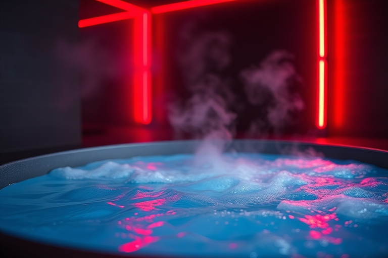
Red light therapy works through photobiomodulation, a process where photons at specific wavelengths (630 to 660 nm red, 810 to 850 nm near-infrared) are absorbed by cytochrome c oxidase in the mitochondria, displacing inhibitory nitric oxide, increasing ATP production by up to 50%, triggering beneficial reactive oxygen species signaling, and activating transcription factors that drive cellular repair, anti-inflammatory responses, and tissue regeneration. This isn't a theoretical model. It's a well-characterized biochemical pathway supported by thousands of peer-reviewed studies.
The Mitochondrial Mechanism
To understand red light therapy, you need to understand how mitochondria produce energy. The electron transport chain (ETC) is a series of protein complexes (I through V) embedded in the inner mitochondrial membrane. Electrons pass through these complexes, driving protons across the membrane to create a gradient. Complex V (ATP synthase) uses this gradient to produce ATP.
Cytochrome C Oxidase (Complex IV)
Complex IV, cytochrome c oxidase (CCO), is the terminal enzyme in the ETC. It accepts electrons from cytochrome c and transfers them to molecular oxygen, producing water. This step is rate-limiting for the entire chain. If Complex IV slows down, the entire energy production system backs up.
CCO contains copper and heme centers that absorb light in specific wavelength bands. The absorption peaks align precisely with the wavelengths used in red light therapy: 630 to 660 nm (red) and 810 to 850 nm (near-infrared). This isn't coincidence. The therapeutic wavelengths were identified by mapping the action spectrum of photobiomodulation against the absorption spectrum of CCO.
Nitric Oxide Inhibition and Release
Under conditions of cellular stress, inflammation, or hypoxia, nitric oxide (NO) binds to the copper and heme centers of CCO, competing with oxygen for the binding site. When NO occupies these sites, electron flow through Complex IV slows, ATP production decreases, and excess electrons leak to form damaging superoxide radicals.
Red and NIR photons displace NO from CCO through a photodissociation mechanism. The photon energy breaks the NO-metal bond, releasing NO and restoring oxygen binding. This has two consequences:
- ATP production resumes: With oxygen properly bound, electron flow normalizes and ATP output increases. Studies show up to 50% increases in ATP production in treated cells.
- Free NO signals vasodilation: The released NO enters surrounding tissue and acts as a vasodilator, increasing blood flow to the treated area. This enhances oxygen and nutrient delivery and supports the removal of metabolic waste.
Reactive Oxygen Species Signaling
The increase in electron flow through the ETC produces a brief, mild increase in reactive oxygen species (ROS). This sounds harmful, but at low levels, ROS function as signaling molecules rather than damaging agents. This is another example of hormesis: a small dose of a potentially harmful stimulus triggers a beneficial adaptive response.
View data table
| Category | Evidence and Efficacy Score | Detail |
|---|---|---|
| Cold Exposure | 72 | Metabolic activation |
| Red Light | 65 | Mitochondrial support |
| CGM Tracking | 82 | Glucose optimization |
| Peptide Stacks | 70 | Targeted protocols |
| Nootropics | 55 | Cognitive enhancement |
Transcription Factor Activation
The ROS pulse activates several transcription factors:
- NF-kB: At low activation levels (as from PBM), NF-kB promotes cell survival, proliferation, and anti-inflammatory gene expression. This contrasts with chronic, high-level NF-kB activation from disease, which promotes inflammation. The dose determines the outcome.
- AP-1 (activator protein 1): Promotes collagen gene expression, which explains the skin rejuvenation effects of red light therapy.
- Nrf2: Activates the antioxidant response element (ARE), upregulating endogenous antioxidant enzymes (SOD, catalase, glutathione peroxidase). This makes cells more resilient to future oxidative stress.
Light Penetration and the Optical Window
The body isn't transparent to light. Different wavelengths are absorbed, scattered, or reflected by different tissue components:
Check your GLP-1 eligibility
Use our free BMI Calculator to see if you may qualify for provider-reviewed GLP-1 therapy.
Try the BMI Calculator →- Hemoglobin absorbs strongly below 600 nm, blocking blue, green, and yellow light from penetrating deeply.
- Water absorbs strongly above 1,100 nm, blocking far-infrared and longer wavelengths.
- Melanin absorbs broadly across the visible spectrum but less at longer wavelengths.
This creates an "optical window" between approximately 600 and 1,100 nm where light can penetrate tissue most effectively. Within this window, two absorption peaks of CCO create the two therapeutic bands: 630 to 660 nm and 810 to 850 nm.
Penetration Depth
- Red light (630 to 660 nm): Penetrates 2 to 3 mm into tissue. Reaches the dermis, superficial blood vessels, and surface-level nerve endings. Optimal for skin, superficial wounds, and inflammation.
- Near-infrared (810 to 850 nm): Penetrates 3 to 5 cm. Reaches muscle tissue, joint spaces, bone surfaces, and even brain tissue through the skull (though significantly attenuated). Optimal for musculoskeletal conditions, deep tissue recovery, and transcranial applications.
The Biphasic Dose Response (Arndt-Schulz Law)
Photobiomodulation follows a biphasic dose-response pattern. This is critically important for practical application:
- Too little light: Below the threshold dose, no measurable biological effect occurs.
- Optimal dose: Within the therapeutic window, cellular responses are maximized. For most tissues, this falls between 1 and 10 J/cm2 (joules per square centimeter).
- Too much light: Above the optimal dose, the beneficial effects diminish and can reverse. Excessive ROS production shifts from signaling to damage. Cellular function is inhibited rather than stimulated.
This is why more isn't always better with red light therapy. Doubling your treatment time or standing closer doesn't double the benefit. It may reduce it.
Calculating Your Dose
Energy density (J/cm2) = Irradiance (mW/cm2) x Time (seconds) / 1,000
Example: A panel delivering 100 mW/cm2 at your treatment distance, used for 10 minutes (600 seconds):
100 x 600 / 1,000 = 60 J/cm2
This is above the optimal range for most superficial applications but appropriate for deeper tissues at distance (where actual tissue irradiance is lower than surface irradiance due to absorption and scattering).
Cellular and Tissue Effects
Collagen Synthesis
Red light stimulates fibroblast proliferation and upregulates procollagen gene expression through AP-1 activation. Fibroblasts are the primary collagen-producing cells in the dermis. Multiple RCTs have demonstrated measurably increased collagen density after 8 to 12 weeks of red light treatment.
Anti-Inflammatory Effects
PBM reduces inflammatory markers through several mechanisms:
- Decreased prostaglandin E2 (PGE2) production
- Reduced cyclooxygenase-2 (COX-2) expression
- Lowered pro-inflammatory cytokines (TNF-alpha, IL-1beta, IL-6)
- Increased anti-inflammatory cytokine IL-10
Neurological Effects
Near-infrared light applied transcranially reaches cortical neurons and enhances their mitochondrial function. This increases ATP availability for neuronal processes, improves cerebral blood flow through NO-mediated vasodilation, and upregulates BDNF expression. The 40 Hz pulsed frequency appears particularly effective for cognitive applications, potentially through entrainment of gamma oscillations associated with attention and memory. red light therapy benefits complete guide.
Stem Cell Activation
Red and NIR light stimulate mesenchymal stem cell proliferation and differentiation. This has implications for tissue repair throughout the body, from bone healing to cartilage regeneration. The mechanism involves increased mitochondrial activity in stem cells, which provides the energy needed for division and differentiation.
What the Meta-Analyses Show
Several systematic reviews and meta-analyses have evaluated the evidence for PBM:
- Skin rejuvenation: Meta-analyses confirm that PBM improves skin complexion, reduces wrinkle severity, and increases collagen density. Effect sizes are modest but statistically significant.
- Musculoskeletal pain: Strong evidence for osteoarthritis pain reduction. Moderate evidence for tendinopathy and lower back pain. Effect sizes comparable to NSAIDs for some conditions.
- Exercise recovery: Meta-analysis of 46 studies found that PBM applied before or after exercise significantly reduced muscle damage markers and improved recovery.
- Wound healing: Strong evidence for accelerated wound healing, particularly in chronic wounds and diabetic ulcers.
Limitations of the Evidence
Transparency requires acknowledging weaknesses in the research base:
- Dosing inconsistency: Studies use widely varying wavelengths, irradiance levels, treatment times, and frequencies. This makes direct comparisons difficult.
- Small sample sizes: Many trials have fewer than 50 participants.
- Publication bias: Positive results are more likely to be published, potentially inflating perceived efficacy.
- Mechanism specificity: While the CCO mechanism is well-established in vitro, the exact dose reaching deep tissues in vivo is harder to measure and may vary significantly between individuals based on skin pigmentation, body composition, and tissue type.
Despite these limitations, the overall evidence base is large (thousands of studies), the mechanism is biologically plausible, and the safety profile is excellent.
Frequently Asked Questions
- Is red light therapy actually proven to work?
- Yes, for specific applications. The mechanism (photon absorption by cytochrome c oxidase leading to increased ATP) is well-characterized. Clinical evidence is strongest for skin rejuvenation, wound healing, musculoskeletal pain, and exercise recovery. Evidence is growing for cognitive benefits and hormonal support. Not every marketed claim is supported, which is why understanding the science helps you evaluate specific applications critically.
- Why do specific wavelengths matter?
- Cytochrome c oxidase has specific absorption peaks at approximately 630-660 nm and 810-850 nm. Light at other wavelengths is either not absorbed by CCO (no photobiomodulation effect) or is absorbed by other tissue components (hemoglobin, water) before reaching the mitochondria. The wavelength determines whether photons reach and interact with the target enzyme.
- Can red light therapy cause cancer by stimulating cell growth?
- This is a theoretical concern that hasn't been validated in research. PBM stimulates normal cellular processes (ATP production, repair mechanisms) rather than promoting uncontrolled growth. But as a precaution, most practitioners advise against treating directly over known tumors. The photon energies used in PBM (1.4 to 2.0 eV) are far below the threshold for ionizing radiation and can't damage DNA directly.
- Does skin color affect red light therapy effectiveness?
- Melanin absorbs visible light, so darker skin pigmentation does reduce the amount of red light reaching deeper tissues. Near-infrared (810-850 nm) is less affected by melanin than red (660 nm). People with darker skin may benefit from slightly longer treatment times or closer treatment distances, though clinical significance of this difference needs more research.
- How is red light therapy different from laser therapy?
- Both use the same wavelengths and the same mechanism (photobiomodulation). The difference is the light source. Lasers produce coherent, collimated light. LEDs produce incoherent, divergent light. Research has shown that the biological effects are similar when the same wavelength and energy density reach the target tissue. LEDs are cheaper, safer (no risk of retinal damage from coherent beams), and can cover larger treatment areas.
See your options in about 2 minutes
Take the free quiz and see what fits you. Quick, private, and no commitment to continue.
See my options →