Executive Summary

Key Takeaways
- Dihexa is a synthetic angiotensin IV analog that potentiates HGF/c-Met signaling to drive synaptogenesis at extraordinarily low concentrations
- Animal studies demonstrate reversal of age-related and chemically induced cognitive deficits with oral administration at 2 mg/kg/day
- The compound is orally bioavailable, crosses the blood-brain barrier, and has a prolonged circulating half-life of approximately 12.68 days (IV administration)
- Fosgonimeton (ATH-1017), a prodrug of dihexa, has completed Phase 2/3 trials for Alzheimer's disease with mixed results
- Serious theoretical safety concerns exist regarding cancer promotion via c-Met pathway activation, with zero long-term safety data in humans
Dihexa (N-hexanoic-Tyr-Ile-(6) aminohexanoic amide) is a synthetic hexapeptide analog of angiotensin IV that has generated extraordinary interest in neuroscience and the nootropic community due to its reported ability to drive synapse formation at concentrations roughly seven orders of magnitude lower than brain-derived neurotrophic factor (BDNF). Developed at Washington State University by Dr. Joseph Harding and colleagues, dihexa represents a fundamentally different approach to cognitive enhancement - one that targets the hepatocyte growth factor (HGF)/c-Met receptor system rather than traditional neurotransmitter pathways.
The story of dihexa begins with decades of research into the renin-angiotensin system (RAS), specifically the observation that angiotensin IV, a metabolic fragment of angiotensin II, possessed unexpected procognitive properties when administered to laboratory animals. Through systematic chemical modification aimed at improving metabolic stability and oral bioavailability, Harding's team produced a series of angiotensin IV analogs, with dihexa emerging as the most potent and pharmacologically promising candidate. Unlike its parent peptide, dihexa resists enzymatic degradation, crosses the blood-brain barrier efficiently, and can be administered orally - a rare combination for peptide-based compounds.
What sets dihexa apart from virtually every other nootropic compound is its mechanism of action. Rather than modulating acetylcholine, dopamine, glutamate, or other neurotransmitters, dihexa works by binding to hepatocyte growth factor and potentiating its interaction with the c-Met receptor tyrosine kinase. This triggers downstream signaling cascades, primarily through PI3K/Akt and MAPK/ERK pathways, that promote the formation of new synaptic connections (synaptogenesis), increase dendritic spine density (spinogenesis), and support neuronal survival. In essence, dihexa doesn't just make existing neural circuits work harder or faster; it appears to build new structural connections between neurons.
The preclinical evidence for dihexa's cognitive-enhancing properties is striking. In aged rats with natural cognitive decline, oral dihexa at 2 mg/kg per day restored performance in the Morris water maze to levels indistinguishable from healthy young controls. In scopolamine-induced amnesia models, dihexa reversed memory deficits with similar efficacy. And in APP/PS1 transgenic mice - the most widely used genetic model of Alzheimer's disease - dihexa rescued spatial learning and memory while simultaneously reducing neuroinflammation, decreasing pro-inflammatory cytokines IL-1beta and TNF-alpha, and increasing the anti-inflammatory cytokine IL-10 (McCoy AT, et al. Journal of Pharmacology and Experimental Therapeutics. 2013;345(2):284-294).
The compound's pharmaceutical lineage extends to fosgonimeton (ATH-1017), a phosphate prodrug of dihexa developed by Athira Pharma that has progressed through multiple clinical trials for Alzheimer's disease and Parkinson's disease. While the Phase 2/3 LIFT-AD trial did not meet its primary endpoint, secondary analyses and earlier Phase 2 SHAPE trial data showed directional improvements in cognitive scores and biomarkers, keeping the HGF/c-Met approach alive as a therapeutic target.
Yet dihexa also carries serious and largely uncharacterized risks. The HGF/c-Met signaling pathway is a well-established driver of cancer biology. Multiple pharmaceutical companies have developed c-Met inhibitors precisely because excessive c-Met signaling promotes tumor cell proliferation, survival, invasion, and metastasis. No long-term carcinogenicity studies have been conducted with dihexa. No human pharmacokinetic or safety data exist outside of the fosgonimeton clinical program. And a key 2014 mechanistic study was retracted due to data fabrication, weakening portions of the published evidence base. For individuals exploring cognitive enhancement through peptide research, understanding both the promise and the significant unknowns surrounding dihexa is essential.
This report provides a thorough examination of dihexa's discovery, molecular pharmacology, preclinical evidence, comparative potency, oral bioavailability, and safety profile. Every claim is grounded in peer-reviewed research, and every limitation is stated plainly. The goal is to give researchers, clinicians, and informed individuals the most complete picture available of what dihexa can and cannot do, what we know and what remains dangerously uncertain.
Key Takeaways
- Dihexa is a synthetic angiotensin IV analog that potentiates HGF/c-Met signaling to drive synaptogenesis at extraordinarily low concentrations
- Animal studies demonstrate reversal of age-related and chemically induced cognitive deficits with oral administration at 2 mg/kg/day
- The compound is orally bioavailable, crosses the blood-brain barrier, and has a prolonged circulating half-life of approximately 12.68 days (IV administration)
- Fosgonimeton (ATH-1017), a prodrug of dihexa, has completed Phase 2/3 trials for Alzheimer's disease with mixed results
- Serious theoretical safety concerns exist regarding cancer promotion via c-Met pathway activation, with zero long-term safety data in humans
- Dihexa is not FDA-approved for any indication and remains a research compound with no established human dosing protocol
Discovery from Angiotensin IV Research
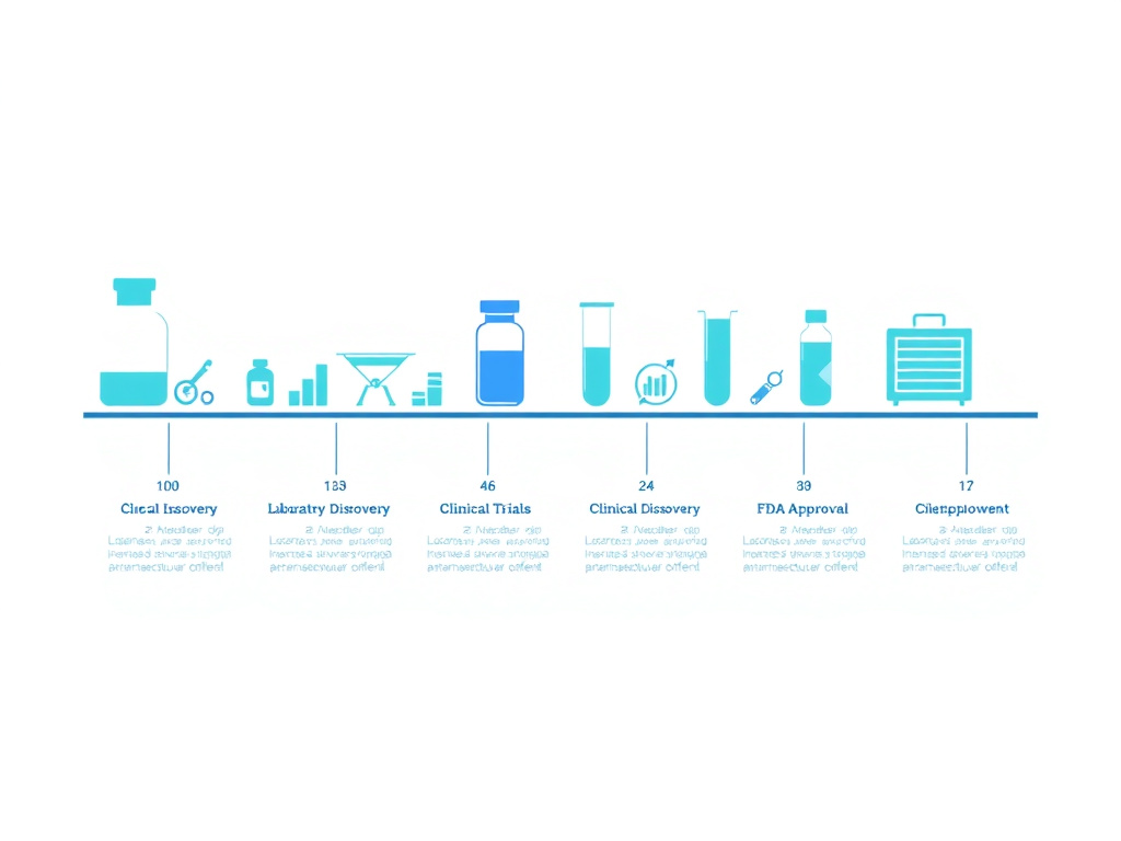
Figure 2: Historical timeline of angiotensin IV receptor research and the systematic development of metabolically stabilized analogs leading to dihexa
The discovery of dihexa did not happen in a single flash of insight. It emerged from over two decades of systematic research into a corner of the renin-angiotensin system that most cardiovascular researchers had overlooked - the biological activity of angiotensin IV, a six-amino-acid fragment once dismissed as an inactive metabolic byproduct.
The Renin-Angiotensin System: Beyond Blood Pressure
The renin-angiotensin system (RAS) has been studied since the late 19th century, primarily in the context of blood pressure regulation and fluid homeostasis. The canonical pathway is well known: renin cleaves angiotensinogen to produce angiotensin I, which angiotensin-converting enzyme (ACE) then converts to angiotensin II. Angiotensin II acts on AT1 and AT2 receptors to regulate vasoconstriction, aldosterone secretion, and sympathetic nervous system activity. ACE inhibitors and angiotensin receptor blockers (ARBs) that target this pathway remain among the most prescribed medications worldwide.
But angiotensin II doesn't simply bind its receptors and disappear. It undergoes further enzymatic processing. Aminopeptidase A removes the N-terminal aspartate to produce angiotensin III (a heptapeptide). Aminopeptidase N then clips off the arginine to produce angiotensin IV (Val-Tyr-Ile-His-Pro-Phe), a hexapeptide. For decades, these downstream metabolites were considered biologically inert debris - the cellular equivalent of exhaust fumes from the main reaction.
That assumption began to unravel in the late 1980s and early 1990s when several research groups, most prominently the laboratory of John W. Wright and Joseph W. Harding at Washington State University, reported that angiotensin IV had distinct biological effects that could not be explained by activity at AT1 or AT2 receptors. When injected directly into the brains of rats, angiotensin IV improved performance on learning and memory tasks. This was unexpected. The parent molecule angiotensin II had no such effect, and the standard AT1/AT2 receptor framework offered no explanation for why a metabolic fragment would enhance cognition (Braszko JJ, et al. Neuroscience. 1988;27(3):777-783).
Identification of the AT4 Receptor
In 1992, Harding's laboratory identified a specific, high-affinity binding site for angiotensin IV that was pharmacologically distinct from AT1 and AT2 receptors. They designated it the AT4 receptor. This binding site was found in particularly high concentrations in brain regions associated with learning and memory - the hippocampus, neocortex, basal nucleus of Meynert, and cerebellum. The distribution pattern suggested that whatever the AT4 receptor was doing, it was intimately connected to cognitive processing (Harding JW, et al. Brain Research. 1992;583(1-2):340-345).
The identity of the AT4 receptor became a subject of intense investigation and some controversy. In 2001, Albiston and colleagues reported that the AT4 receptor was actually insulin-regulated aminopeptidase (IRAP), a zinc metalloprotease expressed on the surface of neurons and other cell types. Under this model, angiotensin IV enhanced cognition by inhibiting IRAP's enzymatic activity, thereby extending the half-life of neuropeptides like vasopressin, oxytocin, and somatostatin that IRAP normally degrades (Albiston AL, et al. Journal of Biological Chemistry. 2001;276(52):48623-48626).
The IRAP hypothesis was elegant and generated a significant body of supporting research. But the Harding laboratory had been pursuing a parallel line of investigation that would ultimately point in a different direction - toward the hepatocyte growth factor system.
Systematic Development of Metabolically Stable Analogs
A fundamental problem with using angiotensin IV as a cognitive enhancer was its pharmacokinetic profile. As a natural peptide, it was rapidly degraded by aminopeptidases and other proteases in the blood and brain tissue. Its half-life was measured in minutes, it could not cross the blood-brain barrier effectively, and oral administration was impossible because gastrointestinal enzymes would destroy it before absorption.
Starting in the late 1990s, Harding and Wright's team embarked on a systematic medicinal chemistry campaign to create angiotensin IV analogs with improved stability and bioavailability. The strategy involved modifying both the N-terminus and C-terminus of the hexapeptide to block the enzymatic cleavage sites while preserving the core pharmacological activity. Key modifications included replacing the N-terminal valine with a hexanoic acid group (to resist aminopeptidases) and substituting the C-terminal proline-phenylalanine with a 6-aminohexanoic amide (to resist carboxypeptidases).
This work produced a series of compounds designated Norleual (Nle1-AngIV), followed by increasingly optimized analogs. Each generation was tested for binding affinity, metabolic stability, blood-brain barrier penetration, and cognitive effects in animal models. The team evaluated dozens of modifications, mapping out structure-activity relationships with painstaking thoroughness (Benoist CC, et al. Journal of Pharmacology and Experimental Therapeutics. 2011;339(1):35-44).
The Emergence of Dihexa
Dihexa (N-hexanoic-Tyr-Ile-(6) aminohexanoic amide) emerged from this optimization process as the standout compound. Its chemical structure retains the Tyr-Ile core of angiotensin IV - the minimum pharmacophore needed for biological activity - flanked by synthetic modifications that confer extraordinary metabolic stability. The N-terminal hexanoic acid group and C-terminal aminohexanoic amide create a molecule that is resistant to virtually all known peptidases, has increased lipophilicity (which aids blood-brain barrier penetration), and maintains the ability to engage the target binding site with high affinity.
The pharmacokinetic properties of dihexa were remarkable for a peptide-derived compound. Cursory characterization revealed stability in serum, a prolonged circulating half-life of 12.68 days following intravenous administration and 8.83 days following intraperitoneal injection, and confirmed penetration across the blood-brain barrier. Phase I metabolism was extremely low, with an average intrinsic clearance of 2.72 microliters/min/mg and an average microsomal half-life of 509.4 minutes (McCoy AT, et al. Journal of Pharmacology and Experimental Therapeutics. 2013;345(2):284-294).
Perhaps most significant from a drug development perspective was the prediction - later confirmed experimentally - that dihexa was orally bioavailable. The compound's predicted intestinal permeability (Peff value of 1.78) placed it between enalapril (1.25) and piroxicam (2.14), two well-established oral drugs. This meant that unlike virtually every other peptide-based nootropic, dihexa could potentially be taken as a pill rather than requiring injection.
A Surprising Mechanism: The HGF Connection
As dihexa and related analogs were being developed, the Harding laboratory made a discovery that reframed the entire field. While investigating how angiotensin IV analogs produced their cognitive effects, they found that the procognitive and synaptogenic activities of these compounds did not depend on binding to IRAP or any of the classical angiotensin receptor subtypes. Instead, the active compounds shared structural homology with the dimerization domain of hepatocyte growth factor (HGF).
Further experiments demonstrated that dihexa and its relatives bound directly to HGF with high affinity and facilitated HGF's interaction with its receptor, the c-Met tyrosine kinase. When the c-Met system was blocked pharmacologically or genetically, the cognitive benefits of dihexa disappeared entirely. When HGF was absent, dihexa had no effect. The compound was not a standalone agonist; it was an allosteric potentiator of HGF, making the endogenous growth factor more effective at activating its receptor (McCoy AT, et al. Journal of Pharmacology and Experimental Therapeutics. 2013;345(2):284-294).
This was a profound shift. It meant that the cognitive enhancement produced by angiotensin IV and its analogs was not really about the renin-angiotensin system at all. The RAS connection was an accident of molecular evolution - a structural similarity between angiotensin IV and a portion of HGF that allowed the peptide to engage a completely different signaling system. The true target was the HGF/c-Met pathway, one of the most powerful growth factor systems in the body, with roles in embryonic development, tissue regeneration, and - critically - the formation and maintenance of synaptic connections in the brain.
From Bench to Broader Interest
The publication of dihexa's mechanism and preclinical data attracted attention from multiple directions. Athira Pharma (initially Aralez Bio) licensed technology related to the HGF/c-Met approach and developed fosgonimeton (ATH-1017), a phosphate prodrug designed for subcutaneous injection, which entered clinical trials for Alzheimer's and Parkinson's disease. Meanwhile, the compound gained significant popularity in the nootropic and biohacking communities, where its reported potency relative to BDNF generated enormous interest. Those exploring biohacking approaches and cognitive optimization began discussing dihexa alongside more established compounds like semax and selank.
The research peptide market responded accordingly. Dihexa became available from multiple suppliers as a research chemical, typically sold as a lyophilized powder for reconstitution. It's worth understanding that this availability does not imply any regulatory approval. Dihexa has never been approved by the FDA or any other regulatory body for any therapeutic indication. It exists in a regulatory gray zone common to many research peptides, sold explicitly for in vitro and animal research purposes.
The journey from angiotensin IV to dihexa illustrates how basic science can lead to unexpected therapeutic targets. What began as an investigation into a supposedly inactive peptide fragment led to the identification of a new receptor system, the development of a novel chemical scaffold, and ultimately the recognition that one of the body's most powerful growth factor pathways could be harnessed for cognitive enhancement. Whether that harnessing can be done safely remains the central unanswered question.
Timeline of Key Discoveries
- 1988: Braszko et al. report that angiotensin IV improves learning and memory in rats
- 1992: Harding laboratory identifies the AT4 receptor binding site in brain tissue
- 2001: Albiston et al. identify AT4 receptor as insulin-regulated aminopeptidase (IRAP)
- 2004-2011: Systematic development of metabolically stabilized angiotensin IV analogs
- 2013: McCoy et al. publish dihexa's mechanism of action through HGF/c-Met potentiation
- 2014: Benoist et al. confirm procognitive effects depend on HGF/c-Met activation
- 2019-2024: Athira Pharma conducts clinical trials with fosgonimeton (ATH-1017)
- 2021: Chinese researchers demonstrate dihexa rescues cognition in APP/PS1 Alzheimer's mice via PI3K/AKT
HGF/c-Met Signaling Pathway
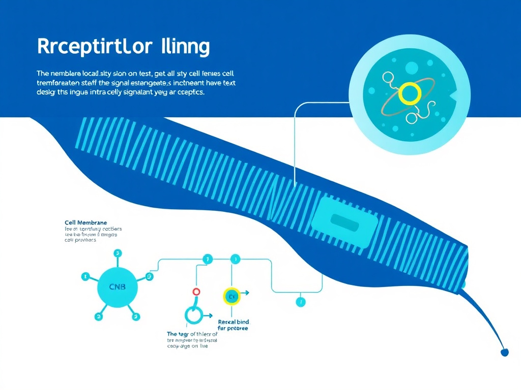
The hepatocyte growth factor (HGF) and its receptor c-Met form one of the most powerful signaling systems in mammalian biology. Originally identified for its role in liver regeneration and embryonic development, the HGF/c-Met axis has since been recognized as a critical regulator of neuronal survival, axonal growth, synaptic formation, and cognitive function. Understanding this pathway at the molecular level is essential to appreciating both why dihexa produces such dramatic effects in preclinical models and why those same effects raise legitimate safety concerns.
Hepatocyte Growth Factor: Structure and Activation
Hepatocyte growth factor is a large, multidomain protein produced primarily by mesenchymal cells - fibroblasts, smooth muscle cells, and certain immune cells. In the brain, HGF is secreted by neurons, astrocytes, and microglia, with particularly high expression in the hippocampus, cortex, and cerebellum. The protein is synthesized as an inactive single-chain precursor (pro-HGF) that requires proteolytic cleavage to become biologically active. This cleavage, carried out by serine proteases including hepatocyte growth factor activator (HGFA) and matriptase, produces a two-chain heterodimer linked by a disulfide bond.
The active HGF molecule consists of an alpha chain containing an N-terminal hairpin domain and four kringle domains, and a shorter beta chain containing a serine protease-like domain that lacks enzymatic activity. The three-dimensional structure of HGF is critical to its function: the N-terminal domain and first kringle domain (NK1 fragment) contain the primary c-Met binding sites, while the remaining kringle domains mediate interactions with heparan sulfate proteoglycans on cell surfaces, concentrating HGF near its target receptors.
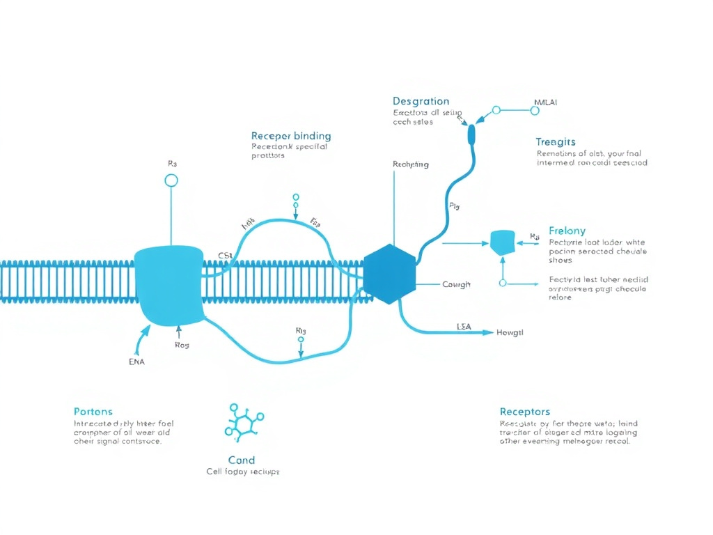
A key structural feature of HGF that connects directly to dihexa's mechanism is the dimerization domain. For full c-Met activation, HGF molecules must form dimers or oligomers that bring two c-Met receptor molecules into close proximity. This receptor dimerization is the triggering event for intracellular signaling. And it is precisely at this dimerization step that dihexa exerts its effect. The peptide binds to HGF in a region that facilitates the formation of active HGF dimers, essentially making the endogenous growth factor more efficient at assembling the molecular complex needed to activate c-Met (McCoy AT, et al. Journal of Pharmacology and Experimental Therapeutics. 2013;345(2):284-294).
The c-Met Receptor: Architecture and Activation
c-Met (also known as MET or hepatocyte growth factor receptor) is a receptor tyrosine kinase encoded by the MET proto-oncogene on chromosome 7. The mature receptor is a disulfide-linked heterodimer consisting of a 50-kDa alpha subunit (entirely extracellular) and a 145-kDa beta subunit that spans the cell membrane and contains the intracellular kinase domain.
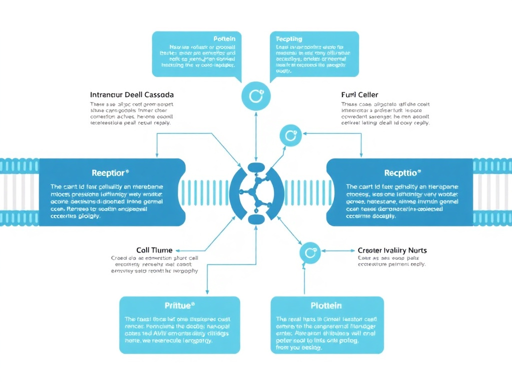
When HGF binds to c-Met and induces receptor dimerization, the intracellular kinase domains of the two receptor molecules undergo transphosphorylation. The critical initial phosphorylation events occur at tyrosine residues 1234 and 1235 (Y1234 and Y1235) within the activation loop of the kinase domain. This activates the intrinsic kinase activity of c-Met, which then phosphorylates additional tyrosine residues - most critically Y1349 and Y1356 near the C-terminus. These phosphotyrosines create a multisubstrate docking site that serves as a platform for recruiting and activating a constellation of downstream signaling molecules.
The docking site at Y1349/Y1356 is unusual among receptor tyrosine kinases in its ability to bind a remarkably diverse set of signaling adaptors and enzymes. These include Grb2 (growth factor receptor-bound protein 2), Gab1 (Grb2-associated binder 1), PI3K (phosphoinositide 3-kinase), phospholipase C-gamma (PLC-gamma), Shp2 (SH2 domain-containing phosphatase 2), Src family kinases, and STAT3 (signal transducer and activator of transcription 3). This multi-pathway engagement explains why HGF/c-Met signaling produces such broad biological effects - a single receptor activation event simultaneously triggers multiple intracellular cascades.
PI3K/Akt Pathway: Neuronal Survival and Growth
The PI3K/Akt pathway is arguably the most important downstream effector of c-Met signaling in the context of cognitive enhancement. When PI3K is recruited to the activated c-Met receptor complex (primarily through Gab1), it phosphorylates membrane phospholipids to generate phosphatidylinositol-3,4,5-trisphosphate (PIP3). PIP3 then recruits Akt (also called protein kinase B) to the membrane, where it is phosphorylated and activated by PDK1 and mTORC2.
Activated Akt phosphorylates dozens of downstream substrates that collectively promote neuronal survival, growth, and plasticity. Key targets include:
- GSK-3beta: Akt phosphorylates and inactivates glycogen synthase kinase-3beta, removing a brake on multiple growth-promoting pathways and stabilizing proteins involved in cytoskeletal remodeling and synaptic function
- mTORC1: Through phosphorylation of TSC2, Akt activates the mechanistic target of rapamycin complex 1, which drives protein synthesis necessary for new synapse construction and long-term memory consolidation
- BAD and caspase-9: Akt phosphorylates pro-apoptotic proteins, suppressing programmed cell death and promoting neuronal survival under stress conditions
- CREB: Through intermediate kinases, Akt can activate cyclic AMP response element-binding protein, a transcription factor that drives expression of BDNF, synaptic proteins, and other genes critical for long-term plasticity
- FOXO transcription factors: Akt phosphorylates and inactivates FOXO proteins, which otherwise promote apoptosis and oxidative stress responses
In the 2021 study by Xie and colleagues using APP/PS1 transgenic mice, dihexa treatment was shown to activate the PI3K/AKT signaling pathway in brain tissue, and this activation was directly linked to reduced neuronal apoptosis, decreased neuroinflammation, and improved cognitive performance. When PI3K was inhibited pharmacologically, the neuroprotective and anti-inflammatory effects of dihexa were abolished, confirming that the PI3K/Akt cascade is a necessary mediator of dihexa's benefits (Xie Y, et al. Brain Sciences. 2021;11(11):1487).
MAPK/ERK Pathway: Synaptic Plasticity and Memory
The mitogen-activated protein kinase (MAPK) cascade, specifically the Ras-Raf-MEK-ERK pathway, is the second major signaling arm activated by c-Met. This pathway is recruited through Grb2, which binds to phosphorylated c-Met and activates the Ras GTPase through the guanine nucleotide exchange factor SOS (Son of Sevenless).
Active Ras triggers a phosphorylation cascade: Ras activates Raf kinases, which phosphorylate and activate MEK1/2, which in turn phosphorylates and activates ERK1/2 (extracellular signal-regulated kinases). ERK1/2 then phosphorylates a wide range of cytoplasmic and nuclear targets that regulate gene expression, protein synthesis, and cytoskeletal dynamics.
In neurons, the MAPK/ERK pathway is critical for:
- Long-term potentiation (LTP): ERK activation is required for the late phase of LTP, the sustained strengthening of synaptic connections that underlies long-term memory formation. Blocking ERK prevents the conversion of short-term memory to long-term memory
- Dendritic spine remodeling: ERK regulates actin dynamics and the activity of spine-associated Rho GTPases that control the size, shape, and stability of dendritic spines - the postsynaptic structures where excitatory synapses are located
- Synaptic protein expression: ERK-driven transcription increases production of synaptic scaffolding proteins (PSD-95, Homer, Shank), neurotransmitter receptors (AMPA, NMDA subunits), and vesicle-associated proteins that strengthen and maintain synaptic connections
- BDNF expression: ERK activates CREB, which drives transcription of BDNF, creating a positive feedback loop where HGF/c-Met activation leads to increased BDNF production, further supporting synaptic plasticity
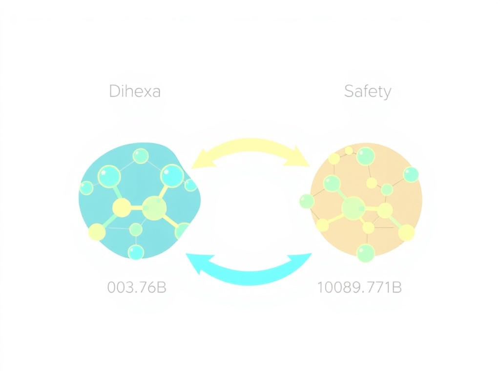
This connection to BDNF is particularly relevant to the widely cited claim that dihexa is "ten million times more potent than BDNF." The comparison originated from in vitro assays measuring synaptogenic activity at different molar concentrations. Because dihexa works upstream - by amplifying HGF/c-Met signaling, which in turn increases BDNF expression and activates overlapping downstream pathways - extremely small concentrations of dihexa can trigger a cascade that ultimately produces effects comparable to much larger amounts of directly applied BDNF. The potency comparison is technically accurate at the assay level but can be misleading if interpreted as meaning dihexa is millions of times more effective at improving cognition overall.
PLC-gamma and Calcium Signaling
A third signaling branch activated by c-Met involves phospholipase C-gamma (PLC-gamma). When recruited to the activated receptor, PLC-gamma hydrolyzes phosphatidylinositol-4,5-bisphosphate (PIP2) into two second messengers: inositol-1,4,5-trisphosphate (IP3) and diacylglycerol (DAG). IP3 triggers calcium release from the endoplasmic reticulum, while DAG activates protein kinase C (PKC). Both calcium and PKC are important regulators of synaptic transmission, neurotransmitter release, and plasticity.
In hippocampal neurons, HGF-induced PLC-gamma activation promotes neurite outgrowth through PKC-dependent pathways and enhances calcium-dependent processes involved in synaptic vesicle fusion and neurotransmitter release. This provides an additional mechanism by which dihexa, through HGF potentiation, could enhance both synaptic formation and synaptic transmission (Royal I, et al. Molecular Biology of the Cell. 2000;11(5):1709-1727).
STAT3 Pathway: Neuroprotection and Anti-inflammation
c-Met also activates the STAT3 (signal transducer and activator of transcription 3) pathway. STAT3 is phosphorylated either directly by c-Met or indirectly through Src family kinases, then dimerizes and translocates to the nucleus where it functions as a transcription factor. In the context of neurodegeneration, STAT3 activation promotes expression of anti-apoptotic genes (Bcl-2, Bcl-xL, Mcl-1) and anti-inflammatory mediators.
The anti-inflammatory effects observed in the APP/PS1 mouse studies - reduced IL-1beta, reduced TNF-alpha, increased IL-10 - likely involve STAT3-mediated transcriptional regulation in microglia and astrocytes, suggesting that dihexa's benefits extend beyond direct neuronal effects to include modulation of the neuroinflammatory environment. For researchers interested in the intersection of inflammation and neurodegeneration, compounds like BPC-157 and thymosin alpha-1 work through related but distinct anti-inflammatory mechanisms.
How Dihexa Specifically Engages This System
Dihexa does not bind c-Met directly. It does not replace HGF. And it does not activate c-Met signaling in the absence of endogenous HGF. Instead, dihexa functions as an allosteric potentiator of HGF. The compound binds to HGF protein and facilitates the formation of the active HGF dimer that is required for efficient c-Met activation.
This mechanism has important implications. First, dihexa's effects are dependent on the presence of endogenous HGF. In brain regions with low HGF expression, dihexa would be expected to have minimal effects. In regions with high HGF expression - the hippocampus, for example - dihexa amplifies an existing signal rather than creating a new one. Second, the potentiator mechanism means that dihexa's effects scale with HGF levels, which change in response to injury, aging, and disease. In conditions where HGF levels are elevated (such as after brain injury or in certain neurodegenerative states), dihexa could produce disproportionately strong effects.
Third, and critically for safety assessment, the potentiator mechanism means that dihexa enhances c-Met activation wherever HGF is present - not just in the brain. HGF and c-Met are widely expressed throughout the body, including in the liver, kidneys, lungs, and gastrointestinal tract. They are also expressed in many tumor types, where c-Met signaling drives cancer cell proliferation and metastasis. This systemic exposure to enhanced c-Met signaling is the primary basis for concerns about dihexa's long-term safety, a topic examined in detail in the safety section of this report.
Pathway Summary
Dihexa potentiates HGF → HGF activates c-Met → c-Met phosphorylation recruits Gab1, Grb2, PI3K, PLC-gamma, STAT3 → Parallel activation of PI3K/Akt (survival, growth), MAPK/ERK (plasticity, gene expression), PLC-gamma/PKC/Ca2+ (transmission, outgrowth), and STAT3 (neuroprotection) → Net result: increased synaptogenesis, spinogenesis, neuronal survival, and reduced neuroinflammation
Synaptic Connectivity Enhancement
Synaptic connectivity is the physical foundation of cognition. Every memory, every learned skill, every cognitive process depends on the precise pattern and strength of connections between neurons. Dihexa's most remarkable preclinical property is its apparent ability to drive the formation of new synaptic connections - synaptogenesis - at concentrations far below those required by any other known compound. This section examines the cellular and molecular details of how dihexa remodels neural circuits.
The Biology of Synapse Formation
Synapse formation is an enormously complex process that requires coordinated action between the presynaptic neuron (which will release neurotransmitters) and the postsynaptic neuron (which will receive the signal). The process begins with axonal growth - the extension of a presynaptic axon toward a potential postsynaptic partner. Growth cones at the tips of extending axons use molecular guidance cues to navigate toward appropriate targets.
Once contact is made, a series of molecular events converts the initial contact into a functional synapse. On the presynaptic side, vesicle release machinery must be assembled, including synaptobrevin, syntaxin, SNAP-25 (the SNARE complex), synaptotagmin (the calcium sensor), and the vesicle recycling apparatus. On the postsynaptic side, neurotransmitter receptors must be clustered, scaffolding proteins (PSD-95, Homer, Shank) must be organized, and the actin cytoskeleton must be remodeled to form or enlarge dendritic spines.
In the adult brain, synapse formation is more constrained than during development but still occurs, particularly in the hippocampus and other regions involved in learning. Adult synaptogenesis is regulated by neurotrophic factors (BDNF, NGF, NT-3), growth factors (HGF, EGF, FGF), and activity-dependent signals (calcium influx through NMDA receptors, activation of CaMKII). The rate of new synapse formation typically declines with age, contributing to age-related cognitive decline.
Dihexa's Synaptogenic Potency
The most widely cited property of dihexa is its synaptogenic potency. In assays measuring the ability of compounds to increase the number of functional synaptic connections between cultured hippocampal neurons, dihexa produced measurable effects at picomolar concentrations (10^-12 M). By comparison, BDNF required nanomolar to low micromolar concentrations (10^-9 to 10^-6 M) to produce equivalent synaptogenic effects. This represents a roughly 10^7-fold (ten million-fold) difference in molar potency.
It is important to understand what this comparison does and does not mean. The potency difference reflects the fact that dihexa works by a catalytic mechanism - it potentiates an endogenous growth factor - rather than acting as a direct trophic factor itself. A single dihexa molecule, by facilitating HGF dimerization, can trigger a c-Met signaling cascade that produces sustained downstream effects. BDNF, by contrast, acts as a stoichiometric ligand - each molecule activates a single TrkB receptor, and the duration of signaling depends on receptor occupancy.
The amplification cascade works something like this: one dihexa molecule facilitates the activation of HGF. One active HGF dimer activates two c-Met receptor molecules. Each activated c-Met recruits multiple signaling adaptors (Gab1, Grb2, PI3K, etc.). Each adaptor activates multiple downstream kinases (Akt, ERK, PKC). Each kinase phosphorylates multiple substrates. The net result is an enormous amplification of signal from a very small initial input. This is fundamentally different from BDNF/TrkB signaling, where the amplification is more modest because the initial receptor activation event requires direct ligand binding at much higher concentrations.
Spinogenesis: Building New Dendritic Spines
Dendritic spines are small protrusions from the dendrite surface where the majority of excitatory synapses are located. They come in several morphological types - thin, stubby, mushroom-shaped, and filopodia-like - and their size, shape, and number are closely correlated with synaptic strength and cognitive function. Mushroom spines, with their large heads and narrow necks, represent the most mature and functionally stable synaptic connections. Thin spines are more labile and may represent newly formed or learning-related connections.
In hippocampal neuron cultures treated with dihexa, researchers observed significant increases in the total number of dendritic spines per unit length of dendrite. The spine density increase was dose-dependent and accompanied by increases in the expression of postsynaptic density protein 95 (PSD-95), a scaffolding protein that is a reliable marker of functional excitatory synapses. Increased PSD-95 clustering was confirmed by immunofluorescence microscopy, showing that the new spines contained organized postsynaptic machinery capable of receiving synaptic input.
The mechanism by which dihexa drives spinogenesis involves HGF/c-Met-dependent activation of Rho family GTPases, particularly Rac1 and Cdc42. These molecular switches control actin polymerization - the fundamental cytoskeletal process that builds and reshapes dendritic spines. Rac1 activation promotes the formation of lamellipodia-like structures that serve as precursors for new spines, while Cdc42 drives filopodia extension, the initial protrusive event in spine formation. Both processes are downstream of PI3K/Akt signaling activated through c-Met.
Synaptophysin and Presynaptic Remodeling
Synapse formation requires coordinated changes on both sides of the synaptic cleft. In the APP/PS1 mouse study (Xie et al., 2021), dihexa treatment increased the expression of synaptophysin (SYP), a presynaptic vesicle protein that serves as a marker for functional presynaptic terminals. Synaptophysin is a component of the synaptic vesicle membrane and is involved in vesicle docking, fusion, and recycling. Its upregulation indicates not just that new postsynaptic spines are being formed, but that matching presynaptic terminals are being assembled to create complete, functional synaptic connections.
The coordination between pre- and postsynaptic remodeling suggests that dihexa-driven HGF/c-Met signaling activates trans-synaptic signaling pathways. These likely include the neurexin-neuroligin system (which physically bridges pre- and postsynaptic membranes and coordinates their maturation), the ephrin-Eph receptor system (which regulates contact-dependent synaptic signaling), and secreted factors like Wnt proteins and TGF-beta family members that are released by neurons to promote synapse maturation in their local environment.
Long-Term Potentiation Enhancement
Long-term potentiation (LTP) is the sustained strengthening of synaptic transmission that occurs when two neurons are repeatedly activated together. It is the leading cellular model for learning and memory. LTP has two phases: early LTP (lasting 1-3 hours), which depends on post-translational modifications of existing proteins, and late LTP (lasting hours to days), which requires new protein synthesis and structural changes at synapses.
HGF/c-Met signaling, as potentiated by dihexa, is positioned to enhance both phases of LTP. The rapid activation of PI3K/Akt and MAPK/ERK cascades can modulate AMPA receptor trafficking (inserting more receptors into the postsynaptic membrane to strengthen transmission) within minutes - supporting early LTP. The transcriptional effects mediated through CREB, STAT3, and mTORC1-dependent translational control drive production of new synaptic proteins needed for late LTP and for converting newly formed connections into stable, lasting ones.
Electrophysiological studies in brain slices from dihexa-treated animals have shown enhanced LTP in the hippocampal CA1 region, the brain area most critical for spatial memory and the first region affected in Alzheimer's disease. The enhancement was not just a modest increase in LTP magnitude but a qualitative improvement - treated animals showed LTP that was both larger in amplitude and more resistant to reversal (depotentiation) than controls.
Network-Level Effects and Circuit Remodeling
Individual synapse formation is important, but cognition depends on the organized patterns of connectivity across neural networks. A key question about dihexa is whether the new synapses it promotes are functionally appropriate - connecting the right neurons in the right patterns - or whether they represent disorganized overgrowth that could actually impair function.
The available evidence, while limited, suggests that dihexa-driven synaptogenesis is guided by the same activity-dependent rules that normally govern circuit refinement. Because dihexa is a potentiator rather than an agonist, it amplifies signaling that is already occurring at sites of active HGF/c-Met engagement. In the brain, HGF expression is upregulated at sites of activity and in response to learning-related stimuli. This means that dihexa should preferentially enhance synapse formation at active circuits - the circuits being used for learning - rather than promoting random connectivity throughout the brain.
However, this remains partially theoretical. No studies have directly mapped the connectivity patterns of newly formed synapses in dihexa-treated brains using modern connectomics techniques. The behavioral data (improved performance on specific cognitive tasks) is consistent with appropriate circuit enhancement, but more detailed anatomical and functional circuit analysis is needed. For those interested in other peptides that support neural health, semax and selank have been studied for their neuroprotective properties through different mechanisms, while P21 is another peptide with neurogenic properties that researchers have explored for cognitive applications.
Comparison to Other Synaptogenic Approaches
Several other approaches to promoting synaptogenesis exist, and comparing them to dihexa illuminates its unique position. BDNF, delivered directly or upregulated through exercise and environmental enrichment, promotes synaptogenesis through TrkB receptor activation. The effects are well-characterized but limited by BDNF's poor pharmacokinetics (it doesn't cross the blood-brain barrier and is rapidly degraded). CJC-1295/Ipamorelin combinations promote growth hormone release, which supports general brain health but through indirect mechanisms.
IGF-1 and its analogs, including IGF-1 LR3, promote neuronal survival and have some synaptogenic activity, but primarily through the PI3K/Akt pathway without the HGF-specific signaling. The nootropic racetam compounds (piracetam, aniracetam) modulate AMPA receptor kinetics and may have mild synaptogenic effects, but at potencies orders of magnitude below dihexa. NAD+ and its precursors support mitochondrial function and sirtuin activity, promoting neuronal health through metabolic pathways rather than growth factor signaling.
Dihexa stands alone in terms of raw synaptogenic potency per mole of compound applied. But potency is not the same as efficacy or safety. The question of whether more synaptogenesis is always better - and at what point enhanced connectivity becomes maladaptive - remains unresolved.
Critical Distinction: Potency vs. Clinical Effect
The "10 million times more potent than BDNF" claim refers specifically to the molar concentration required to produce measurable synaptogenic effects in cell culture assays. It does not mean dihexa is 10 million times better at improving cognition, 10 million times safer, or 10 million times more effective as a treatment. The potency comparison reflects the amplification cascade inherent in dihexa's mechanism (potentiating a growth factor rather than acting as one directly) and should not be extrapolated to clinical effectiveness without appropriate caveats.
Animal Cognitive Studies
The preclinical evidence for dihexa's cognitive-enhancing effects comes from a series of animal studies conducted primarily at Washington State University and subsequently confirmed by independent research groups in China and elsewhere. These studies employed multiple cognitive paradigms, different routes of administration, various models of cognitive impairment, and both young and aged animals. The consistency of positive results across these varied experimental conditions constitutes the strongest argument for dihexa's biological activity.
Scopolamine-Induced Amnesia Model
Scopolamine is a muscarinic acetylcholine receptor antagonist that produces reversible amnesia when administered to rodents. It is one of the most widely used pharmacological models for testing potential cognitive enhancers because it mimics, in a simplified way, the cholinergic deficit that characterizes Alzheimer's disease. Animals treated with scopolamine show impaired performance on spatial memory tasks, object recognition, and contextual fear conditioning.
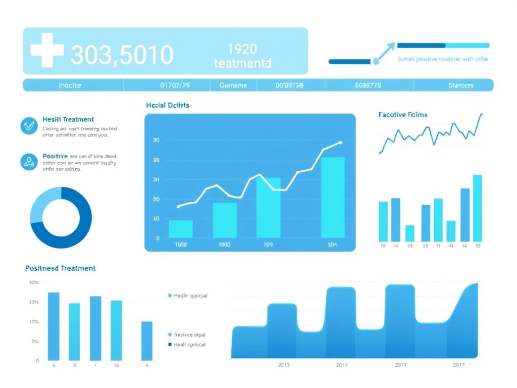
In the foundational dihexa study (McCoy et al., 2013), rats were treated with scopolamine (0.5 mg/kg, intraperitoneally) to induce cognitive impairment, then given dihexa (2 mg/kg/day, orally by gavage) or vehicle control. Cognitive performance was assessed using the Morris water maze, a spatial learning task where rats must learn the location of a hidden platform in a circular pool of opaque water by using visual landmarks around the room.
The results were striking. Scopolamine-treated rats showed severe impairment in learning the platform location, making many more errors and taking significantly longer to find the platform compared to untreated controls. Scopolamine-treated rats that also received dihexa, however, performed nearly identically to healthy controls. The compound reversed approximately 90% of the scopolamine-induced deficit, restoring correct response rates from approximately 35% (scopolamine alone) to approximately 80% (scopolamine plus dihexa), compared to 85% for untreated controls.
Morris Water Maze Performance
Data adapted from McCoy AT, et al. J Pharmacol Exp Ther. 2013;345(2):284-294.
On probe trials - where the platform is removed and the time spent searching in the correct quadrant is measured as an index of memory retention - dihexa-treated animals showed strong spatial memory, spending significantly more time in the target quadrant than chance level (25%) and not significantly different from healthy controls. This indicated that dihexa didn't just improve procedural aspects of the task (swimming speed, search strategy) but genuinely restored the ability to form and retain spatial memories.
Age-Related Cognitive Decline Model
Perhaps more relevant to real-world cognitive enhancement than pharmacological amnesia models is the effect of dihexa on naturally occurring age-related cognitive decline. Aged rats (typically 22-24 months old, equivalent to roughly 65-75 human years) show well-documented deficits in spatial learning, working memory, and cognitive flexibility compared to young adults (3-6 months old).
When aged rats were treated with oral dihexa at 2 mg/kg/day for 14 days, their performance on the Morris water maze improved to levels statistically indistinguishable from young adult controls. This was not a subtle effect. Aged control rats showed the typical pattern of slow learning, inconsistent performance, and poor retention on probe trials. Aged rats receiving dihexa showed learning curves that overlapped with those of young adults - they found the platform faster, made fewer errors across training trials, and showed strong spatial memory on probe tests.
Chronic administration studies extending to 28 days revealed that the cognitive enhancement was sustained without evidence of tolerance development. Animals continued to perform well throughout the treatment period, and some studies reported that benefits persisted for a period after treatment cessation, consistent with the structural nature of the changes (new synapses, once formed and stabilized, would be expected to persist beyond the period of pharmacological intervention).
Histological analysis of hippocampal tissue from aged dihexa-treated rats showed increased dendritic spine density, elevated expression of synaptic markers (PSD-95, synaptophysin), and evidence of enhanced neurogenesis in the dentate gyrus subgranular zone. These structural changes provided a plausible anatomical basis for the observed cognitive improvements.
APP/PS1 Transgenic Mouse Model of Alzheimer's Disease
The APP/PS1 mouse model carries human transgenes for amyloid precursor protein (APP) with the Swedish mutation and presenilin-1 (PS1) with the deltaE9 mutation. These mice develop progressive amyloid-beta plaque deposition, neuroinflammation, synaptic loss, and cognitive deficits that recapitulate key features of Alzheimer's disease. They are among the most widely used preclinical models for testing potential Alzheimer's therapies.
In 2021, Xie and colleagues published a study in Brain Sciences examining dihexa's effects in APP/PS1 mice at 6 months of age (when cognitive deficits are clearly established but not yet at end-stage severity). Mice received dihexa by intraperitoneal injection for 28 days, then underwent behavioral testing followed by detailed histological and molecular analyses (Xie Y, et al. Brain Sciences. 2021;11(11):1487).
Key findings from this study included:
- Cognitive restoration: Dihexa restored spatial learning and memory in the Morris water maze to near-wildtype levels. Treated APP/PS1 mice showed significantly reduced escape latencies during training and increased time in the target quadrant on probe trials
- Neuronal preservation: Nissl staining revealed increased neuronal cell counts in the hippocampus of dihexa-treated versus vehicle-treated APP/PS1 mice, suggesting reduced neuronal loss
- Synaptic marker restoration: Synaptophysin (SYP) protein expression was significantly increased in treated mice, indicating preservation or restoration of presynaptic terminals
- Anti-inflammatory effects: Dihexa markedly reduced activation of astrocytes (GFAP staining) and microglia (Iba1 staining) in the hippocampus. Pro-inflammatory cytokines IL-1beta and TNF-alpha were significantly decreased, while the anti-inflammatory cytokine IL-10 was significantly increased
- PI3K/Akt pathway activation: Western blot analysis confirmed increased phosphorylation of Akt (p-Akt/Akt ratio) in the brains of dihexa-treated mice. When the PI3K inhibitor LY294002 was co-administered, it blocked dihexa's neuroprotective and anti-inflammatory effects, confirming the causal role of PI3K/Akt signaling
This study was significant for several reasons. It used a genetic model of Alzheimer's disease rather than a pharmacological impairment model, providing evidence relevant to actual neurodegenerative pathology. It identified a specific intracellular signaling mechanism (PI3K/Akt) that could be targeted for therapeutic development. And it demonstrated anti-inflammatory effects that extend beyond simple synaptic enhancement, suggesting that dihexa might address multiple pathological processes contributing to cognitive decline. For those tracking developments in Alzheimer's therapeutics, the GLP-1 research hub covers the emerging connection between metabolic signaling and neurodegeneration, as semaglutide and other GLP-1 agonists have also shown neuroprotective properties in preclinical models.
Novel Object Recognition
Beyond the Morris water maze, dihexa has been tested in the novel object recognition (NOR) paradigm. In this test, animals are first exposed to two identical objects, then after a delay, one object is replaced with a novel one. Normal animals preferentially explore the novel object, reflecting their memory of the familiar one. Animals with memory impairments show no preference, exploring both objects equally.
Young healthy rats given acute doses of dihexa (0.5-2 mg/kg by oral gavage) showed enhanced novel object recognition compared to vehicle-treated controls, spending significantly more time exploring the novel object at both short (1-hour) and long (24-hour) retention intervals. This suggested that dihexa improved both short-term and long-term recognition memory, consistent with effects on both early and late phases of memory consolidation.
Contextual Fear Conditioning
Contextual fear conditioning tests the ability of animals to associate an environment (context) with an aversive stimulus (typically a mild foot shock). When returned to the same context, animals that have formed the association show freezing behavior - a natural defensive response. The hippocampus is critical for contextual (as opposed to cued) fear conditioning, making this a valuable assay for hippocampus-dependent memory.
Dihexa-treated animals showed enhanced contextual fear conditioning, with increased freezing responses when returned to the training context. The effect was dose-dependent and present at oral doses as low as 0.5 mg/kg. This provided converging evidence from a third behavioral paradigm that dihexa enhances hippocampus-dependent memory formation.
Traumatic Brain Injury Model
Recent preclinical work has extended dihexa testing to models of traumatic brain injury (TBI). A 2025 preprint from the Harding laboratory described the effects of dihexa in rats subjected to repetitive mild TBI (a model relevant to sports-related concussions and military blast injuries). This study represented the first evidence that a small molecule activator of the HGF/MET system can improve cognitive function following repeated mild TBI. Treated rats showed ameliorated deficits in working memory performance compared to TBI-injured vehicle controls, suggesting potential applications beyond age-related decline and Alzheimer's disease.
Route of Administration Comparisons
Multiple administration routes have been tested in animal studies:
| Route | Effective Doses | Onset | Key Findings |
|---|---|---|---|
| Oral (gavage) | 0.5-2 mg/kg/day | Days 3-5 of dosing | Confirmed oral bioavailability; most extensively tested route |
| Intraperitoneal (IP) | 0.1-1 mg/kg/day | Days 1-3 of dosing | Faster onset than oral; used in APP/PS1 studies |
| Intracerebroventricular (ICV) | 0.01-0.1 nmol | Within hours | Extremely low doses effective; confirms central mechanism |
| Subcutaneous (SC) | 0.5-2 mg/kg/day | Days 2-4 of dosing | Comparable efficacy to oral; sustained release kinetics |
The fact that intracerebroventricular administration (which delivers the compound directly into the brain ventricles, bypassing systemic exposure) was effective at extremely low doses confirmed that dihexa's cognitive effects are mediated through central nervous system actions rather than peripheral mechanisms. The efficacy of oral administration, while requiring higher doses due to gastrointestinal and hepatic first-pass effects, validated the compound's potential utility as a practical therapeutic agent.
Limitations of the Preclinical Data
While the consistency of positive results across multiple models and paradigms is encouraging, several important limitations must be acknowledged. The total number of published studies is relatively small - perhaps a dozen papers from a handful of research groups. Most of the original work comes from a single laboratory (Harding/Wright at Washington State University), and while independent confirmation from Chinese research groups strengthens the findings, broader replication is needed.
The retraction of a 2014 paper from the Harding laboratory due to data fabrication (specifically, manipulation of blot images) raises legitimate concerns about data integrity, even though the retracted study was not the primary source of the cognitive enhancement data. Additionally, no published study has conducted a full dose-response analysis with multiple endpoints, and long-term toxicology data are entirely absent from the preclinical literature.
Finally, the translation from animal cognitive assays to human cognitive enhancement is notoriously unreliable. Many compounds that dramatically improve rodent performance on the Morris water maze have failed completely in human clinical trials. The fosgonimeton (ATH-1017) clinical trial results - where the drug failed to meet primary endpoints despite strong preclinical data - illustrate this translational gap directly.
Potency Comparison to Other Nootropics
The nootropic landscape spans everything from caffeine and L-theanine to sophisticated peptide therapeutics, and positioning dihexa within this spectrum requires careful attention to what "potency" means in different contexts. Dihexa's synaptogenic potency is unmatched. But synaptogenic potency is not the same as cognitive improvement magnitude, safety margin, or practical utility. This section compares dihexa to the major categories of cognitive-enhancing compounds.
Dihexa vs. BDNF: The Ten-Million-Fold Claim
The most commonly cited comparison - that dihexa is approximately seven orders of magnitude (10 million times) more potent than BDNF at promoting synaptogenesis - deserves careful unpacking. This figure comes from in vitro assays where hippocampal neurons were treated with varying concentrations of either dihexa or BDNF, and synapse formation was quantified by counting co-localized presynaptic (synapsin-1 or synaptophysin) and postsynaptic (PSD-95 or Homer) puncta along dendrites.
In these assays, dihexa produced statistically significant increases in synapse number at concentrations as low as 10^-12 M (picomolar). BDNF required concentrations of 10^-5 to 10^-4 M (tens of micromolar) to produce comparable synaptogenic effects. The concentration ratio gives the roughly 10^7-fold potency difference.
But there are critical caveats. First, the assays measured a single endpoint (synapse count) under specific in vitro conditions. In vivo, where pharmacokinetics, tissue distribution, receptor availability, and competing signals all come into play, the potency difference would be expected to narrow considerably. Second, BDNF has many biological effects beyond synaptogenesis - it promotes neuronal survival, modulates neurotransmitter release, and regulates ion channel expression - that are not captured by a synapse-counting assay. Third, dihexa requires the presence of endogenous HGF to function, while BDNF acts directly on its receptor. In a system with low HGF levels, dihexa might have reduced efficacy regardless of its intrinsic potency.
Dihexa vs. Peptide Nootropics
Within the peptide nootropic category, dihexa occupies a unique position due to its growth factor-potentiating mechanism. Other peptide nootropics work through very different pathways:
| Compound | Primary Mechanism | Oral Bioavailability | Human Data | Safety Profile |
|---|---|---|---|---|
| Dihexa | HGF/c-Met potentiation | Yes (confirmed in animals) | None (prodrug ATH-1017 in trials) | Largely unknown; cancer risk concern |
| Semax | BDNF/NGF upregulation, melanocortin receptors | Intranasal only | Approved in Russia since 1994 | Generally well-tolerated; decades of clinical use |
| Selank | GABA modulation, tuftsin analog | Intranasal only | Approved in Russia since 2009 | Anxiolytic; well-characterized safety |
| P21 | CNTF mimetic, neurogenesis | Intranasal | Limited | Preclinical only; unclear long-term |
| Pinealon | Gene expression regulation (CNS) | Yes (tripeptide) | Limited Russian studies | Generally considered safe; short peptide |
| Epithalon | Telomerase activation, pineal regulation | No (injectable) | Limited Russian studies | Limited data; theoretical concerns similar to dihexa |
Semax presents perhaps the most interesting comparison. Like dihexa, semax increases BDNF expression in the hippocampus and has documented nootropic effects. But semax works through melanocortin receptor modulation and direct neurotrophic factor upregulation rather than growth factor potentiation. It has decades of clinical use in Russia, with a well-characterized safety profile, though it must be administered intranasally and cannot be taken orally. Its effects are more modest than dihexa's in preclinical models, but its safety record is incomparably better documented.
Selank, a tuftsin analog, provides cognitive benefits primarily through anxiolytic and GABA-modulating effects rather than structural synaptogenesis. It represents a fundamentally different approach - optimizing cognitive function by reducing anxiety and improving mood state, rather than building new synaptic connections. For individuals whose cognitive difficulties stem from stress or anxiety, selank's mechanism might be more appropriate than dihexa's growth factor approach.
Dihexa vs. Racetams and Small-Molecule Nootropics
The racetam family (piracetam, aniracetam, oxiracetam, pramiracetam, phenylpiracetam) represents the original synthetic nootropic compounds. They primarily modulate AMPA receptor kinetics, increasing receptor sensitivity to glutamate and thereby enhancing excitatory synaptic transmission. Some racetams also increase cerebral blood flow and have mild effects on acetylcholine and dopamine systems.
Racetams operate at an entirely different biological level than dihexa. They optimize the function of existing synapses (making transmission more efficient) rather than creating new ones. Their effects are modest by comparison - typical improvements of 10-20% on specific cognitive measures in human studies - but they have decades of safety data in humans and are generally well-tolerated. Piracetam has been available since the 1960s with an extensive clinical record.
Modafinil, another frequently discussed nootropic, promotes wakefulness and cognitive performance primarily through dopamine transporter inhibition and orexin system modulation. Like racetams, it enhances the function of existing circuits rather than building new ones. It is FDA-approved for narcolepsy and has well-characterized pharmacokinetics and side effects in humans.
Dihexa vs. Growth Hormone Secretagogues
Growth hormone secretagogues like CJC-1295/Ipamorelin, MK-677, sermorelin, and tesamorelin promote growth hormone release, which in turn increases circulating IGF-1 levels. IGF-1 crosses the blood-brain barrier and has well-documented neuroprotective and neurotrophic effects, including promotion of hippocampal neurogenesis and synaptic plasticity.
The cognitive benefits of growth hormone secretagogues are indirect and generally modest compared to dihexa's preclinical effects. However, their mechanism is broader - affecting whole-body metabolism, body composition, sleep quality, and recovery in addition to neural function. For individuals seeking general health optimization alongside cognitive support, growth hormone secretagogues may offer a more balanced benefit profile. MK-677 in particular has oral bioavailability and documented effects on sleep architecture that indirectly support cognitive function.
Dihexa vs. Mitochondrial and Longevity Peptides
Within the mitochondrial and longevity peptide category that includes dihexa in the FormBlends peptide research library, several compounds offer cognitive benefits through fundamentally different mechanisms:
SS-31 (elamipretide) targets the inner mitochondrial membrane, stabilizing cardiolipin and improving electron transport chain efficiency. By enhancing mitochondrial function in neurons, SS-31 supports the enormous energy demands of synaptic transmission and maintenance. Its cognitive effects come from optimizing cellular energetics rather than building new connections.
MOTS-c, a mitochondria-derived peptide, activates AMPK signaling and has emerging evidence for neuroprotective effects. Humanin, another mitochondria-derived peptide, protects neurons against amyloid-beta toxicity and oxidative stress. Both work through metabolic and protective mechanisms rather than growth factor potentiation.
NAD+ and its precursors (NMN, NR) support sirtuin activity, DNA repair, and mitochondrial biogenesis. The cognitive benefits of NAD+ supplementation are thought to arise from improved neuronal energy metabolism and reduced age-related DNA damage accumulation. FOXO4-DRI targets senescent cells for clearance, potentially removing dysfunctional cells that contribute to neuroinflammation and cognitive decline.
Summary Potency Table
| Compound | Mechanism Class | Synaptogenic Potency | Human Evidence | Risk Level |
|---|---|---|---|---|
| Dihexa | Growth factor potentiator | Highest known (picomolar) | None direct; prodrug failed trials | High (unknown long-term) |
| BDNF (direct) | Neurotrophin | High (nanomolar) | Not a drug; endogenous | N/A (not pharmacologically viable) |
| Semax | Neurotrophic upregulator | Moderate | Approved in Russia | Low |
| Piracetam | AMPA modulator | Minimal | Extensive (60+ years) | Very low |
| Modafinil | DAT/orexin modulation | None documented | FDA-approved | Low-moderate |
| CJC-1295/Ipamorelin | GH secretagogue | Indirect (via IGF-1) | Clinical data exists | Low-moderate |
| NAD+ | Metabolic cofactor | Indirect (energetics) | Growing human data | Low |
The fundamental tension in evaluating dihexa is between its extraordinary preclinical potency and its almost complete absence of human safety data. Every other compound in this comparison with meaningful human evidence has undergone formal safety evaluation. Dihexa has not. For researchers and individuals weighing cognitive enhancement options, this risk-benefit asymmetry should be the central consideration. The FormBlends science page provides additional context on evidence-based approaches to peptide selection.
Oral Bioavailability
Oral bioavailability is the holy grail of peptide drug development. The vast majority of peptide therapeutics require injection because the gastrointestinal tract destroys them before they can be absorbed. Dihexa's ability to be administered orally and still reach the brain in pharmacologically active concentrations is one of its most distinguishing features, and it results directly from the deliberate chemical modifications that transformed angiotensin IV into a drug-like molecule.
Why Most Peptides Cannot Be Taken Orally
To appreciate why dihexa's oral bioavailability is exceptional, it helps to understand the barriers that defeat most peptide drugs in the gut. The gastrointestinal tract is designed to break down proteins and peptides into individual amino acids for absorption. This destruction occurs at multiple levels: pepsin in the stomach cleaves peptide bonds in an acidic environment; pancreatic proteases (trypsin, chymotrypsin, elastase, carboxypeptidase) attack from multiple angles in the small intestine; brush border peptidases on the surface of enterocytes provide a final line of enzymatic defense.
Even if a peptide survives these enzymes, it must cross the intestinal epithelial barrier. Most peptides are too large and too hydrophilic to diffuse through cell membranes. They are too big for paracellular transport (squeezing between cells). And they typically lack the structural features recognized by active transport systems designed for nutrients and small molecules. The result is that oral bioavailability for most unmodified peptides ranges from 0.1% to less than 2%.
Consider the peptides commonly used for cognitive or metabolic enhancement. BPC-157, while it has some oral activity for gastrointestinal conditions, achieves much higher systemic levels through injection. Semaglutide required decades of development and a specialized absorption enhancer (SNAC) to achieve oral bioavailability, and even then, only about 1% of the oral dose reaches systemic circulation. Most growth hormone secretagogue peptides like sermorelin and CJC-1295/Ipamorelin must be injected subcutaneously.
How Dihexa Achieves Oral Bioavailability
Dihexa overcomes these barriers through its molecular design. The N-terminal hexanoic acid cap and C-terminal aminohexanoic amide modification serve dual purposes: they block the recognition sites for aminopeptidases and carboxypeptidases (protecting against enzymatic degradation), and they increase the molecule's overall lipophilicity (aiding membrane permeation).
The compound's small size also helps. At just three amino acid equivalents with modified termini, dihexa is significantly smaller than most bioactive peptides. Its molecular weight (approximately 507 Da) falls within the range typically considered compatible with oral absorption, and its computed log P (a measure of lipophilicity) places it in the same zone as many orally available small-molecule drugs.
Computational modeling predicted that dihexa would have an effective intestinal permeability (Peff) of 1.78, placing it between two well-established oral drugs: enalapril (Peff = 1.25), an ACE inhibitor taken daily by millions of patients, and piroxicam (Peff = 2.14), a nonsteroidal anti-inflammatory drug. This predicted permeability was consistent with the observed oral efficacy in animal studies, where doses of 0.5-2 mg/kg/day produced significant cognitive enhancement when given by oral gavage (McCoy AT, et al. Journal of Pharmacology and Experimental Therapeutics. 2013;345(2):284-294).
Blood-Brain Barrier Penetration
Reaching the bloodstream is only half the challenge for a CNS-active compound. It must also cross the blood-brain barrier (BBB), a highly selective endothelial barrier that separates circulating blood from brain extracellular fluid. The BBB blocks the passage of most large molecules, charged molecules, and hydrophilic compounds, allowing only small, lipophilic molecules and those recognized by specific transport systems to enter the brain.
Dihexa's increased lipophilicity (resulting from the N-hexanoic acid and aminohexanoic amide modifications) facilitates passive diffusion across the BBB. The compound's relatively small molecular weight keeps it below the typical size threshold for BBB permeation. And unlike many peptides that carry multiple charged amino acid side chains, dihexa's modified structure has reduced overall charge, further aiding membrane crossing.
Direct confirmation of BBB penetration came from pharmacokinetic studies in which dihexa was administered systemically (intravenously or intraperitoneally) and subsequently detected in brain tissue at concentrations consistent with pharmacological activity. The extremely low doses effective when dihexa was injected directly into the brain ventricles (intracerebroventricular administration, at 0.01-0.1 nmol) compared to systemic doses (mg/kg range) provided a rough estimate of BBB penetration efficiency.
Pharmacokinetic Parameters
The pharmacokinetic characterization of dihexa, while described as "cursory" by the original investigators, revealed some remarkable properties:
| Parameter | Value | Significance |
|---|---|---|
| Half-life (IV administration) | 12.68 days | Extraordinarily long for a peptide; suggests extensive protein binding or tissue sequestration |
| Half-life (IP administration) | 8.83 days | Slightly shorter than IV, consistent with first-pass metabolism |
| Intrinsic clearance (Cl_int) | 2.72 microliters/min/mg | Very low, indicating resistance to hepatic metabolism |
| Microsomal half-life | 509.4 minutes (~8.5 hours) | Very stable in liver microsomes; minimal Phase I metabolism |
| Serum stability | Stable | Resistant to circulating peptidases and esterases |
| Predicted Peff (intestinal) | 1.78 | Intermediate; comparable to established oral drugs |
The most striking feature is the circulating half-life of over 12 days following IV administration. For context, the half-life of native angiotensin IV is measured in seconds to minutes. Even semaglutide, which was specifically engineered for extended half-life through albumin binding, has a half-life of approximately 7 days. Dihexa's prolonged persistence in circulation likely results from a combination of its metabolic stability (resistance to enzymatic degradation), lipophilicity-driven tissue distribution (accumulation in adipose tissue and other lipophilic compartments), and possible protein binding (association with albumin or other plasma proteins).
Implications of the Long Half-Life
The extended half-life has both positive and negative implications. On the positive side, it means that infrequent dosing could maintain therapeutic concentrations. In animal studies, once-daily oral dosing was effective, but the pharmacokinetics suggest that every-other-day or even less frequent dosing might be sufficient once steady-state levels are achieved.
On the negative side, the long half-life means that if adverse effects occur, they cannot be quickly reversed by stopping the drug. With a 12-day half-life, approximately 60 days (five half-lives) would be required to eliminate 97% of the compound after the last dose. During that washout period, the effects of dihexa on HGF/c-Met signaling would continue, including any unwanted effects on cell growth or other pathways. This prolonged exposure window significantly increases the risk associated with any toxic or oncogenic effects, because the window of potential harm extends weeks beyond the last administered dose.
For comparison, most cognitive-enhancing drugs have much shorter half-lives that allow rapid washout if problems arise: modafinil (12-15 hours), piracetam (4-5 hours), and even long-acting compounds like methylphenidate (3.5 hours for the active enantiomer). The ability to quickly stop a medication and have it leave the body is an important safety feature that dihexa largely lacks.
Oral Administration Protocols in Research Settings
In published animal studies, oral dihexa has been administered by gavage (direct delivery to the stomach through a tube) at doses of 0.5-2 mg/kg/day, typically in a vehicle of sterile water or saline. The most commonly used effective dose was 2 mg/kg/day for cognitive enhancement in both scopolamine models and aging models.
Because no human clinical trials of dihexa itself (as opposed to the prodrug fosgonimeton) have been conducted, there is no established human oral dose. Simple allometric scaling from rat doses provides only rough estimates, and these should not be used to determine human doses without proper Phase I safety studies. The appropriate human dose, if dihexa is ever tested clinically, would need to be established through formal dose-escalation studies with careful safety monitoring.
For researchers working with dihexa in laboratory settings, the compound's stability in solution is a practical advantage. Unlike many peptides that require reconstitution immediately before use and degrade within hours at room temperature, dihexa's resistance to hydrolysis allows for more practical storage and handling. However, proper cold storage (-20C or colder) and protection from light remain standard recommendations for maintaining compound integrity. Those seeking practical guidance on peptide handling can find general information through the FormBlends dosing calculator, though specific dihexa protocols are not included due to the compound's research-only status.
Oral Bioavailability Comparison
Among cognitive-enhancing peptides, dihexa's oral bioavailability is exceptional. Most nootropic peptides (semax, selank, DSIP) require intranasal or injectable delivery. BPC-157 has some oral activity for GI targets but limited systemic bioavailability. Only MK-677 (ibutamoren, a peptidomimetic GH secretagogue) offers comparable oral bioavailability in the nootropic peptide space, though its mechanism is entirely different.
Safety Concerns & Unknown Risks

Dihexa presents a safety challenge unlike most research peptides. Its mechanism of action - potentiating one of the most powerful growth factor pathways in human biology - raises theoretical risks that are serious, plausible, and almost entirely untested. The absence of long-term safety data is not just a gap in knowledge; it is a gap in knowledge about a pathway known to drive cancer, with a compound that has a half-life measured in days rather than hours. This section lays out what is known, what is unknown, and what the available evidence suggests about the risk landscape.
The Cancer Risk: c-Met as a Proto-Oncogene
The single most significant safety concern surrounding dihexa is its potential to promote cancer through chronic activation of the HGF/c-Met signaling pathway. c-Met is encoded by the MET proto-oncogene, and its dysregulation has been implicated in the development and progression of numerous cancer types including lung cancer, hepatocellular carcinoma, renal cell carcinoma, gastric cancer, colorectal cancer, breast cancer, and glioblastoma.
The evidence linking c-Met to cancer is not theoretical or speculative. It is among the most well-established oncogenic pathways in cancer biology:
- MET gene amplification is a driver mutation in approximately 5-10% of non-small cell lung cancers and is a mechanism of resistance to EGFR-targeted therapies
- MET mutations (including activating mutations in the kinase domain and splice site mutations leading to exon 14 skipping) directly cause constitutive receptor activation and drive tumor growth
- HGF overexpression in the tumor microenvironment promotes invasion, metastasis, and resistance to chemotherapy across multiple cancer types
- c-Met/HGF autocrine loops, where tumor cells produce both HGF and c-Met, create self-sustaining growth signals
The pharmaceutical industry has invested billions of dollars in developing c-Met inhibitors and HGF-neutralizing antibodies specifically because excessive c-Met signaling drives cancer. FDA-approved c-Met pathway inhibitors include capmatinib (Tabrecta) and tepotinib (Tepmetko) for MET exon 14 skipping-mutated non-small cell lung cancer. These drugs work by blocking the same pathway that dihexa activates. The irony is stark: oncologists use drugs that inhibit c-Met to treat cancer, while the nootropic community considers using a drug that activates c-Met to enhance cognition.
Does this mean dihexa will cause cancer? Not necessarily. There are important distinctions between pathological c-Met activation (gene amplification, activating mutations, autocrine HGF production) and the physiological potentiation produced by dihexa. Normal HGF/c-Met signaling occurs throughout life without causing cancer in most people. The question is whether chronic pharmacological enhancement of this signaling by dihexa - at what concentrations, for how long, in which tissues - would cross the threshold from physiological to pathological activation.
No carcinogenicity studies have been conducted with dihexa. No long-term animal studies have evaluated tumor formation. This is not a minor oversight in the data; it is a critical missing piece of information for a compound that directly engages an established oncogenic pathway.
Data Integrity Concerns: The 2014 Retraction
A complicating factor in evaluating dihexa's evidence base is the retraction of a key 2014 study from the Journal of Pharmacology and Experimental Therapeutics. The paper, which provided important mechanistic data on how angiotensin IV-derived peptides depend on HGF/c-Met activation for their procognitive effects, was retracted due to data fabrication, specifically manipulation of Western blot images.
The retraction does not invalidate all of dihexa's preclinical data. The cognitive enhancement results from earlier publications and the independent confirmation from the 2021 Chinese APP/PS1 study stand on their own. But the retraction does undermine confidence in specific mechanistic claims and raises questions about data quality from that research group. In any field where data integrity problems have surfaced, additional skepticism and demand for independent replication are appropriate.
Absence of Human Safety Data
Dihexa itself has never been administered to humans in a controlled clinical trial. The closest human data comes from fosgonimeton (ATH-1017), a phosphate prodrug that is converted to dihexa in the body after subcutaneous injection. Fosgonimeton has been tested in Phase 1, Phase 2 (ACT-AD, SHAPE), and Phase 2/3 (LIFT-AD) clinical trials.
The Phase 1 results indicated that fosgonimeton was generally safe and well-tolerated in the short-term dosing period studied. The Phase 2 and 2/3 trials, while they did not meet their primary efficacy endpoints, provided some safety data over 26-week treatment periods. No dramatic safety signals were reported in the trial publications, but these trials involved relatively small patient populations (hundreds, not thousands) monitored for relatively short durations (months, not years) - insufficient to detect rare events or long-term risks like cancer development, which typically requires years or decades to manifest.
The fosgonimeton trials also involved subcutaneous injection of a prodrug, which produces different pharmacokinetics than oral dihexa. The tissue distribution, peak-to-trough ratios, and duration of c-Met pathway activation may differ substantially between the two formulations. Safety data from fosgonimeton cannot be straightforwardly applied to oral dihexa use.
Potential for Maladaptive Neural Remodeling
Even setting aside cancer risk, there are concerns about whether indiscriminate synaptogenesis could produce negative cognitive effects. The brain's circuits are organized with extraordinary precision, and adding random connections could potentially disrupt established neural networks rather than enhancing them.
Conditions characterized by excessive synaptic density - such as certain forms of autism spectrum disorder, where failure of normal synaptic pruning leads to hyperconnectivity - demonstrate that more synapses are not always better. Epilepsy, where aberrant excitatory connections drive seizure activity, provides another cautionary example. While dihexa appears to promote activity-dependent synaptogenesis (new connections at active circuits) rather than random overgrowth, the long-term consequences of pharmacologically enhanced connectivity in the human brain are unknown.
Drug Interactions and Contraindications
Because no human pharmacology studies have been conducted, drug interactions are completely uncharacterized. Theoretical concerns include:
- Cancer therapeutics: Dihexa would be expected to counteract c-Met inhibitors (capmatinib, tepotinib) and potentially reduce the efficacy of other targeted cancer therapies. Individuals with any cancer history should consider the theoretical risk of growth factor pathway activation very carefully
- Growth factor therapies: Compounds that already increase HGF levels (such as certain stem cell treatments or tissue repair strategies) could produce additive or excessive c-Met activation when combined with dihexa
- Anticoagulants: HGF/c-Met signaling affects endothelial cell function and angiogenesis; potential interactions with anticoagulants or antiplatelet agents are theoretically possible but untested
- Insulin and diabetes medications: The original AT4 receptor was identified as insulin-regulated aminopeptidase (IRAP), and while dihexa's primary mechanism appears to be HGF-mediated rather than IRAP-mediated, residual effects on insulin signaling pathways cannot be ruled out
Regulatory and Legal Status
Dihexa is not FDA-approved for any therapeutic indication. It is not classified as a dietary supplement. It is not a controlled substance in the United States (as of 2026). It is sold by research chemical suppliers as a "research compound" intended for laboratory use only, not for human consumption.
The regulatory landscape for research peptides has been tightening. In 2023, the FDA began more actively enforcing against companies selling peptides for implied human use without approval. While dihexa has not been specifically targeted by enforcement actions, the general regulatory trend suggests that access to research peptides may become more restricted over time. Individuals considering any peptide research should stay informed about current regulatory requirements in their jurisdiction.
Informed Risk Assessment
For individuals considering dihexa for cognitive enhancement research purposes, the risk-benefit calculus is weighted heavily toward caution:
- The potential benefits (improved memory, increased synaptic connectivity, possible neuroprotection) are real but documented only in animal models
- The potential risks (cancer promotion, maladaptive neural remodeling, unknown drug interactions) are theoretical but grounded in well-established biology
- The duration of exposure from a single dose is very long (half-life over 12 days), limiting the ability to quickly discontinue if problems arise
- No established human dosing protocol exists; any dose selection is guesswork
- No monitoring protocols exist for detecting adverse effects specific to chronic HGF/c-Met pathway activation
Individuals with any personal or family history of cancer, particularly cancers where c-Met/HGF signaling is implicated (lung, liver, kidney, stomach, breast, brain), should consider the theoretical risk profile especially carefully. Those interested in evidence-based cognitive enhancement may find better risk-benefit profiles in compounds with established human safety data, such as semax, or in approaches that enhance BDNF through natural means (exercise, which increases BDNF 2-3 fold, and remains the most thoroughly validated cognitive enhancement intervention in humans).
Safety Summary
- Cancer risk: Theoretical but plausible; c-Met is a known oncogene; no carcinogenicity studies conducted
- Human safety data: None for dihexa; limited for prodrug fosgonimeton (short-term, small trials)
- Long half-life: 12+ days means effects persist for weeks after last dose; cannot rapidly discontinue
- Drug interactions: Completely unknown; theoretical conflicts with cancer therapies
- Data integrity: One key mechanistic study retracted for data fabrication
- Regulatory status: Not approved for any use; sold as research compound only
HGF/c-Met Receptor Biology: Why Dihexa's Target Matters
To appreciate both the promise and the risk of dihexa, you need to understand the hepatocyte growth factor (HGF) and its receptor c-Met at a deeper level. This signaling axis is one of the most powerful growth factor systems in the body, with roles in embryonic development, tissue repair, and - critically - cancer biology. Dihexa's potentiation of this pathway is what makes it so effective for synaptogenesis, and also what makes its long-term safety profile genuinely uncertain.
HGF Structure and Processing
Hepatocyte growth factor is a large protein of 728 amino acids, secreted as an inactive single-chain precursor (pro-HGF) that requires extracellular proteolytic cleavage to become biologically active. The cleavage is performed by serine proteases including hepatocyte growth factor activator (HGFA), matriptase, and urokinase-type plasminogen activator (uPA). This two-step activation mechanism provides an important regulatory checkpoint: HGF activity is controlled not just by its expression level but by the availability of activating proteases at the site of action.
The active form of HGF is a heterodimer consisting of an alpha chain (69 kDa) containing four kringle domains and an N-terminal hairpin loop, and a beta chain (34 kDa) containing a serine protease-like domain that has lost its catalytic activity through evolutionary mutation. The alpha chain mediates binding to the c-Met receptor, while the beta chain contributes to receptor dimerization and activation. HGF binds c-Met with a dissociation constant (Kd) of approximately 1-2 nanomolar, reflecting a high-affinity interaction.
Dihexa doesn't bind to c-Met itself. Instead, it binds to HGF and stabilizes the HGF-c-Met complex, reducing the dissociation rate and thereby increasing the effective duration and magnitude of receptor activation. This allosteric potentiation mechanism means that dihexa amplifies existing HGF signaling rather than creating a new signal. In tissues where HGF is present and active, dihexa increases its effects; in tissues without HGF, dihexa has no effect. This context-dependence is important for understanding both the tissue specificity of dihexa's actions and its safety profile.
c-Met Signaling Cascades in Neurons
The c-Met receptor is a receptor tyrosine kinase that, upon HGF binding and dimerization, undergoes autophosphorylation at multiple tyrosine residues in its intracellular domain. The key phosphorylation sites include Y1234/Y1235 in the activation loop (which activates the kinase domain) and Y1349/Y1356 in the C-terminal docking site (which recruits downstream signaling adaptors). The docking site is particularly important because it creates a multi-substrate binding platform that simultaneously activates several signaling cascades:
- PI3K/Akt pathway: The adaptor protein Gab1 is recruited to phosphorylated c-Met and becomes phosphorylated, creating a binding site for the p85 regulatory subunit of PI3K (phosphoinositide 3-kinase). Activated PI3K generates PIP3 at the plasma membrane, recruiting and activating Akt. In neurons, Akt signaling promotes cell survival (by phosphorylating and inactivating the pro-apoptotic proteins Bad and caspase-9), stimulates protein synthesis (through mTOR activation), and drives neurite outgrowth and dendritic spine formation.
- MAPK/ERK pathway: The adaptor proteins Grb2 and SOS are recruited to c-Met, activating the Ras-Raf-MEK-ERK cascade. In neurons, ERK signaling drives gene expression changes essential for long-term synaptic plasticity, including upregulation of activity-regulated cytoskeletal-associated protein (Arc), brain-derived neurotrophic factor (BDNF), and various synaptic adhesion molecules. ERK also phosphorylates CREB, the transcription factor critical for memory consolidation.
- STAT3 pathway: c-Met directly phosphorylates STAT3 (Signal Transducer and Activator of Transcription 3), which dimerizes and translocates to the nucleus to drive transcription of genes involved in cell survival, proliferation, and differentiation. In the nervous system, STAT3 signaling is important for astrocyte function, neuroinflammatory responses, and neural progenitor cell maintenance.
- Rac1/Cdc42 pathway: Through activation of exchange factors for Rho family GTPases, c-Met signaling drives cytoskeletal reorganization that is essential for axon guidance, dendritic branching, and the structural changes underlying synaptogenesis. This pathway is particularly relevant to dihexa's ability to promote new synaptic connections, as the formation of dendritic spines and presynaptic boutons requires precise cytoskeletal remodeling.
The simultaneous activation of these four major pathways is what gives HGF/c-Met signaling its extraordinary potency for neuronal connectivity. Each pathway contributes a different component of the synaptogenesis process: survival (PI3K/Akt), gene expression (MAPK/ERK), glial support (STAT3), and structural remodeling (Rac1/Cdc42). Dihexa, by potentiating HGF's activation of c-Met, amplifies all four cascades simultaneously.
The Synaptogenesis Process at Molecular Resolution
Dihexa's most notable effect in animal studies is the promotion of new synaptic connections. But what does synaptogenesis actually involve at the molecular level, and how does HGF/c-Met signaling drive it?
Synaptogenesis requires coordinated changes in both the presynaptic (axon terminal) and postsynaptic (dendritic spine) neurons, plus involvement of surrounding astrocytes that help stabilize newly formed synapses. The process unfolds in several stages:
Stage 1 - Axon guidance and target recognition: HGF acts as a chemoattractant for growing axons, guiding them toward potential postsynaptic targets. C-Met activation on the growth cone (the motile tip of the growing axon) triggers Rac1-mediated actin polymerization, steering the axon toward the HGF source. This is the same mechanism used during embryonic nervous system development, but in the adult brain it operates at a much smaller scale during synaptic remodeling.
Stage 2 - Contact and adhesion: When a growing axon terminal contacts a dendrite, trans-synaptic adhesion molecules (neurexins on the presynaptic side, neuroligins on the postsynaptic side) form initial contact points. HGF/c-Met signaling upregulates the expression of these adhesion molecules through ERK/CREB-dependent transcription, increasing the probability that initial contacts will be stabilized rather than retracted.
Stage 3 - Presynaptic differentiation: At the contact site, the axon terminal begins to accumulate synaptic vesicle precursors, active zone proteins (like Bassoon and Piccolo), and voltage-gated calcium channels. This process is driven partly by PI3K/Akt-mediated local protein synthesis and partly by retrograde signals from the postsynaptic cell.
Stage 4 - Postsynaptic differentiation: The dendrite forms a specialized protrusion called a dendritic spine, which is enriched in glutamate receptors (AMPA and NMDA receptors), scaffolding proteins (PSD-95, Homer, Shank), and signaling molecules. Spine formation requires Rac1/Cdc42-mediated actin polymerization, which is directly promoted by c-Met signaling. The resulting spine structure provides a compartmentalized signaling domain that can be independently regulated.
Stage 5 - Synaptic maturation and stabilization: Newly formed synapses undergo a maturation process where the initial thin, unstable "filopodia" spines are converted to larger, more stable "mushroom" spines with larger postsynaptic densities. This stabilization requires sustained activity and molecular signals including BDNF (whose expression is upregulated by ERK/CREB signaling from c-Met activation). Astrocytes also contribute by wrapping around the synapse and releasing factors like thrombospondin that promote synaptic stability.
By potentiating HGF/c-Met signaling, dihexa is thought to accelerate and enhance each of these stages, resulting in a net increase in synaptic density. In animal studies, this translated to approximately 30-50% increases in dendritic spine density in the hippocampus, with corresponding improvements in spatial learning and memory tasks.
Dihexa in Neurodegenerative Disease Research
The diseases that destroy cognitive function - Alzheimer's, Parkinson's, vascular dementia, traumatic brain injury - share a common feature: progressive loss of synaptic connections. If dihexa can genuinely promote new synapse formation in the adult brain, its potential applications in neurodegenerative disease are significant. But the gap between animal data and human clinical evidence remains wide, and the risks of chronic HGF/c-Met potentiation add a layer of complexity that pure efficacy considerations don't capture.
Alzheimer's Disease: The Synaptic Loss Hypothesis
Alzheimer's disease is characterized by the progressive accumulation of amyloid-beta plaques and tau neurofibrillary tangles, but the cognitive decline correlates most strongly with synaptic loss rather than plaque burden. Patients in the earliest stages of cognitive impairment already show 25-35% reductions in synaptic density in the hippocampus and prefrontal cortex, and by moderate-stage disease, synaptic loss exceeds 50% in vulnerable brain regions.
The scopolamine-induced cognitive impairment model used in the original dihexa animal studies creates a temporary cholinergic deficit that mimics some aspects of Alzheimer's pathology. Dihexa completely reversed scopolamine-induced deficits in spatial learning and memory at doses as low as 100 picomoles per kilogram administered intracerebrally, or at low milligram-per-kilogram doses administered orally. This potency, approximately 10 million-fold greater than BDNF on a molar basis for pro-cognitive effects, attracted significant attention in the nootropic community.
However, the scopolamine model is a poor proxy for actual Alzheimer's disease. Scopolamine transiently blocks acetylcholine receptors without producing the amyloid pathology, tau accumulation, neuroinflammation, or progressive neuronal death that characterize the disease. In genuine Alzheimer's pathology, the cellular environment may not support new synapse formation even if HGF/c-Met signaling is enhanced. Amyloid-beta oligomers directly disrupt synaptic adhesion molecules, tau pathology impairs axonal transport of synaptic components, and neuroinflammation creates a hostile microenvironment for synaptic remodeling.
More relevant are studies using transgenic Alzheimer's mouse models (APP/PS1, 5xFAD, and similar lines) that develop actual amyloid pathology. Limited data from these models suggest that HGF/c-Met activation can provide neuroprotection and partially restore cognitive function, but the effects are more modest than in the scopolamine model, and the synaptogenic response is blunted by the ongoing neurodegenerative process.
Traumatic Brain Injury: A More Promising Target?
Traumatic brain injury (TBI) may represent a more suitable target for HGF/c-Met potentiation than chronic neurodegenerative disease. After TBI, there is an acute phase of neuronal death and synaptic disruption, followed by a subacute recovery phase where surviving neurons attempt to form new connections and restore circuit function. This post-injury plasticity window represents a period where the brain is actively trying to rebuild synaptic networks, and enhancing HGF/c-Met signaling during this period could theoretically amplify the natural recovery process.
Endogenous HGF levels rise in the brain after TBI, suggesting that this pathway is already part of the natural repair response. Animal studies of TBI have shown that HGF administration during the recovery phase (days 3-14 post-injury) improves functional outcomes including motor coordination, spatial memory, and social behavior. Whether dihexa, as an HGF potentiator rather than a direct HGF replacement, would produce similar benefits depends on whether the post-TBI HGF elevation is sufficient in magnitude for dihexa to amplify.
Comparison with Other Nootropic Peptides
Dihexa occupies a unique niche in the nootropic peptide space, distinguished by its mechanism of action (synaptogenesis via HGF/c-Met) rather than the neurotransmitter modulation or neuroprotection that characterizes most other cognitive-enhancing peptides.
Semax, a synthetic analog of ACTH(4-10), enhances cognition primarily through BDNF upregulation and modulation of serotonergic and dopaminergic neurotransmission. Its effects are faster in onset (noticeable within hours to days) but mechanistically distinct from dihexa's synaptogenic approach. Semax provides acute cognitive enhancement through neurotransmitter optimization, while dihexa theoretically builds new neural hardware over weeks to months. The two approaches are complementary rather than competing: Semax could provide immediate cognitive support while dihexa works on longer-term structural connectivity improvements.
Selank, a synthetic analog of tuftsin, combines anxiolytic effects with cognitive enhancement through GABAergic modulation and BDNF elevation. For individuals whose cognitive impairment is partly driven by anxiety (which impairs prefrontal cortex function and hippocampal memory consolidation), Selank addresses a contributing factor that dihexa does not. Again, the mechanisms are complementary.
Dihexa's uniqueness lies in its structural rather than functional approach to cognitive enhancement. Most nootropics make existing neural circuits work better (by optimizing neurotransmitter levels, enhancing signal-to-noise ratios, or improving neuronal energy metabolism). Dihexa attempts to build more circuits. This structural approach is theoretically more durable - once a new synapse is formed and stabilized, it persists even after the drug is discontinued - but it's also more uncertain in its outcomes and more concerning in its safety implications.
P21, another nootropic peptide that enhances BDNF-dependent neuroplasticity, shares some mechanistic overlap with dihexa in terms of promoting structural neural changes. However, P21 works through the BDNF/TrkB pathway rather than HGF/c-Met, which may carry a different risk profile. BDNF is more selectively expressed in the nervous system than HGF, potentially reducing off-target concerns. See our biohacking research hub for comprehensive comparisons of cognitive enhancement peptides.
Practical Considerations and Honest Risk Assessment
Dihexa is one of the most polarizing compounds in the peptide research community. Its proponents point to its extraordinary potency and unique mechanism; its critics point to the near-complete absence of human data and the legitimate concerns about chronic HGF/c-Met potentiation. An honest assessment requires weighing both sides without exaggeration in either direction.
The Cancer Risk Question
c-Met is classified as a proto-oncogene, and HGF/c-Met signaling is implicated in the development and progression of multiple cancer types including hepatocellular carcinoma, non-small cell lung cancer, gastric cancer, colorectal cancer, renal cell carcinoma, and glioblastoma. In these cancers, c-Met is often overexpressed, constitutively activated, or amplified, driving tumor cell proliferation, survival, migration, and angiogenesis. Multiple c-Met inhibitors (capmatinib, tepotinib, crizotinib) are approved or in clinical development as anticancer drugs.
This creates an obvious concern: if pharmaceutical companies are developing c-Met inhibitors to treat cancer, could a c-Met potentiator like dihexa promote cancer? The answer is not straightforward. HGF/c-Met signaling in normal tissues is tightly regulated by multiple feedback mechanisms, including negative regulators like LRIG1, MIG-6, and the protein phosphatases PTP1B and DEP-1. Dihexa potentiates the existing HGF signal rather than constitutively activating c-Met, meaning the normal regulatory checkpoints should remain intact.
However, "should remain intact" is not the same as "definitely remain intact," and the long-term consequences of chronically amplified HGF/c-Met signaling have not been studied. Even a modest, sustained increase in c-Met activation could theoretically promote the survival and growth of pre-malignant cells that would otherwise be eliminated by the immune system or by apoptosis. This risk is particularly relevant for individuals with a family history of c-Met-driven cancers or those with known pre-malignant conditions.
The practical implication is that anyone considering dihexa should have baseline cancer screening appropriate for their age and risk factors, and should understand that the cancer risk of chronic use is genuinely unknown. The absence of evidence is not evidence of absence, and the mechanistic concern is scientifically legitimate rather than speculative.
The Data Integrity Issue
The retraction of a key mechanistic paper from the Harding laboratory at Washington State University represents a significant blow to the dihexa evidence base. While the core discovery paper (describing dihexa's synthesis and cognitive effects) has not been retracted, the retraction of a supporting study citing data fabrication raises questions about the overall reliability of the research program. This doesn't mean the core findings are necessarily wrong, but it does mean that independent replication by other laboratories is essential before drawing firm conclusions about dihexa's mechanism and efficacy.
As of early 2026, the independent replication data for dihexa remain limited. A small number of studies from laboratories outside the original research group have confirmed aspects of HGF/c-Met potentiation, but the comprehensive characterization of dihexa's cognitive effects has not been independently reproduced. This places dihexa in a different evidence category than peptides like BPC-157 or Semax, which have been studied by multiple independent groups across different countries.
The Long Half-Life Consideration
Dihexa's estimated half-life of 12+ days creates a unique safety consideration. If adverse effects occur, they cannot be quickly reversed by stopping the drug. Most peptides have half-lives measured in minutes to hours, allowing rapid clearance if problems arise. Dihexa's extended duration means that drug levels remain elevated for weeks after the last dose, and any adverse effects would persist throughout this washout period.
The long half-life also means that with repeated dosing, significant drug accumulation occurs. Steady-state concentrations may be substantially higher than single-dose levels, and the full effects of chronic dosing may not be apparent until several weeks into treatment. This has implications for dose titration: starting with the lowest effective dose and increasing gradually is essential, and the interval between dose adjustments should be at least 3-4 weeks to allow steady-state levels to be reached before evaluating the response.
Who Might Reasonably Consider Dihexa
Given the current evidence landscape, the risk-benefit calculation for dihexa varies dramatically depending on the individual's clinical situation:
Potentially reasonable consideration: Individuals with documented cognitive decline from neurodegenerative disease (Alzheimer's, vascular dementia, post-TBI) who have exhausted approved treatment options and are fully informed about the experimental nature and unknown risks of dihexa use. For these individuals, the severe consequences of untreated progressive dementia may justify the uncertain risks of an experimental intervention.
Questionable consideration: Healthy individuals seeking cognitive enhancement for competitive or professional advantage. For these individuals, the risk-benefit ratio is less favorable because the potential harm (cancer promotion, unknown long-term effects) is weighed against enhancement of already-normal function rather than treatment of a serious disease. Other nootropic approaches with better safety data, such as Semax, Selank, or established racetams, may provide a more favorable risk-benefit profile for cognitive enhancement in healthy individuals.
Monitoring If Pursuing Dihexa Research
For individuals who do pursue dihexa use under medical supervision, a comprehensive monitoring protocol is warranted given the unique safety concerns:
- Baseline cancer screening: Age-appropriate cancer screening (colonoscopy, mammography, PSA, etc.) should be current before initiating dihexa. Any personal or family history of c-Met-associated cancers should be carefully evaluated.
- Liver function: ALT, AST, GGT, and bilirubin at baseline and monthly for the first 3 months, then quarterly. HGF is named "hepatocyte growth factor" for a reason - its primary physiological role is in hepatocyte proliferation and liver regeneration. Sustained potentiation of this pathway warrants close liver monitoring.
- Tumor markers: CEA, AFP, and CA 19-9 at baseline and every 6 months. While these markers are imperfect screening tools, significant elevations would warrant further evaluation.
- Cognitive assessment: Formal cognitive testing (MoCA, ADAS-Cog, or computerized cognitive batteries) at baseline and every 3 months to objectively assess whether the compound is producing measurable benefit. Subjective reports of improved cognition are unreliable due to placebo effects and confirmation bias.
- Renal function: Creatinine and eGFR at baseline and quarterly. c-Met signaling is active in renal tubular cells, and the effects of chronic potentiation on kidney function are unknown.
Use our personalized assessment to explore cognitive enhancement approaches that may be appropriate for your specific needs and risk tolerance. The peptide research hub provides broader context on the available evidence for all cognitive peptides.
Pharmacokinetics, Dosing Rationale, and Administration Considerations
Dihexa's pharmacokinetic profile is one of its most distinctive features and one of its most challenging aspects from a safety perspective. Understanding how this compound behaves in the body is essential for anyone evaluating its use.
Oral Bioavailability and Absorption
One of dihexa's most unusual properties for a peptide-derived compound is its oral bioavailability. Most peptides are rapidly degraded by gastric acid and intestinal proteases, requiring subcutaneous or intravenous administration. Dihexa, despite being derived from angiotensin IV (a peptide), has been modified extensively enough that it resists enzymatic degradation in the GI tract and is absorbed across the intestinal epithelium with sufficient bioavailability to produce central nervous system effects after oral dosing.
The structural features that confer oral bioavailability include the N-terminal hexanoyl cap (which blocks aminopeptidase cleavage), the C-terminal aminohexanoic amide (which blocks carboxypeptidase cleavage), and the overall lipophilicity of the molecule (LogP approximately 2.5), which facilitates passive transcellular absorption across the intestinal epithelium. The relatively small molecular weight (approximately 507 Da) also keeps dihexa below the general molecular weight cutoff for oral absorption (approximately 500-700 Da for non-peptide-like molecules).
After oral administration in animal models, dihexa reaches measurable brain concentrations within 30-60 minutes, consistent with blood-brain barrier penetration. The mechanism of BBB crossing is not fully characterized but likely involves a combination of passive diffusion (facilitated by lipophilicity) and potentially active transport through peptide transport systems. Peak brain concentrations occur approximately 2-4 hours after oral dosing.
The Long Half-Life Problem
Dihexa's estimated half-life of 12+ days is extraordinarily long for a peptide-derived compound. For comparison, most therapeutic peptides have half-lives measured in minutes (native GLP-1: 2 minutes) to hours (semaglutide: 165 hours). Dihexa's extended half-life appears to result from tight binding to plasma proteins (reducing renal filtration), resistance to metabolic degradation (due to its structural modifications), and potential sequestration in tissues (particularly the brain, where it may accumulate in neuronal membranes and synaptic spaces).
The long half-life has several practical consequences. First, steady-state concentrations with repeated dosing are reached only after approximately 5 half-lives, meaning 8-10 weeks of daily dosing before the full effect is achieved. This means that dose-finding based on short-term response may be misleading: a dose that seems well-tolerated during the first few weeks could produce excessive effects (or side effects) once steady-state is reached months later.
Second, if adverse effects occur, they persist for weeks to months after discontinuation. There is no antidote or reversal agent for dihexa. If a patient develops a concerning symptom (unexplained liver enzyme elevation, tumor marker change, neurological adverse effect), they cannot quickly eliminate the drug from their system. This irreversibility is fundamentally different from most nootropics and peptides, which clear the system within hours to days.
Third, drug accumulation with chronic dosing could produce tissue concentrations substantially higher than single-dose levels. The cumulative exposure to HGF/c-Met potentiation over months of chronic dosing is an unprecedented pharmacological experiment, and the long-term consequences are genuinely unknown.
Dosing Protocols in Use
Given the absence of formal dose-finding clinical trials, the dosing protocols used by researchers and self-experimenters are extrapolated from animal data and adjusted based on empirical experience. The most common oral dosing range reported in the nootropic community is 10-40 mg per day, though some individuals use lower doses of 5-10 mg. Sublingual administration is sometimes used in an attempt to improve bioavailability and reduce first-pass hepatic metabolism.
The animal studies that demonstrated cognitive enhancement used intracerebroventricular doses of 2 picomoles per kilogram and oral doses of approximately 0.5-2 mg/kg in rats. Allometric scaling from rat to human doses (using body surface area correction) suggests human-equivalent oral doses in the range of 5-20 mg per day for a 70 kg adult. However, allometric scaling is an imperfect method, and the actual optimal human dose could differ substantially in either direction.
Given the long half-life, some practitioners recommend an intermittent dosing approach: 5 days on, 2 days off, or 3 weeks on, 1 week off. The rationale is to reduce cumulative exposure while still maintaining sufficient HGF/c-Met potentiation for cognitive benefit. Whether intermittent dosing actually reduces risk compared to continuous daily dosing is unknown, but the concept is based on the general pharmacological principle that lower cumulative exposure reduces the probability of toxicity.
Sublingual and intranasal administration routes are sometimes used to bypass first-pass hepatic metabolism, potentially allowing lower systemic doses while achieving adequate brain concentrations. Intranasal delivery, in particular, could provide preferential brain exposure through the olfactory and trigeminal nerve pathways, similar to the delivery route used for Selank and Semax. However, dihexa-specific intranasal bioavailability data are not available, and the optimal intranasal dose has not been established.
Visit our biohacking research hub for broader context on nootropic dosing strategies and evidence levels across different cognitive enhancement approaches.
From Angiotensin IV to Dihexa: The Discovery Pipeline and What It Tells Us
Dihexa's development from angiotensin IV research reveals an important story about serendipity in pharmacology and the unexpected connections between blood pressure regulation and cognitive function. Understanding this development path also illuminates why dihexa has the properties it does and why it differs so dramatically from other nootropics.
Angiotensin IV: The Cognitive Blood Pressure Peptide
The renin-angiotensin system (RAS) is primarily known for its role in blood pressure regulation: angiotensin II (Ang II) is a potent vasoconstrictor that raises blood pressure and is the target of ACE inhibitors and angiotensin receptor blockers. But the RAS has an extensive presence in the brain, where angiotensin peptides serve functions beyond blood pressure control.
Angiotensin IV (Ang IV), a hexapeptide fragment of angiotensin II (Val-Tyr-Ile-His-Pro-Phe), was discovered to enhance memory and learning when injected into the brains of rodents. This was an unexpected finding, as the RAS was not thought to have cognitive functions. The researchers at Washington State University, led by Dr. Joseph Harding, identified that Ang IV's cognitive effects were not mediated through the classical angiotensin receptors (AT1 or AT2) but through a distinct binding site that was later identified as insulin-regulated aminopeptidase (IRAP), also designated the AT4 receptor.
However, subsequent research revealed that the cognitive effects of Ang IV could also be mediated through an alternative mechanism: potentiation of hepatocyte growth factor's interaction with the c-Met receptor. This dual mechanism - IRAP inhibition and HGF/c-Met potentiation - set the stage for the development of analogs that could preferentially target one pathway over the other.
The Norleual and Dihexa Design Process
Dr. Harding's laboratory systematically modified the Ang IV structure to create analogs with improved pharmacological properties. The first significant analog was Nle1-Ang IV (Norleual), which replaced the N-terminal valine with norleucine, improving metabolic stability while retaining cognitive-enhancing properties. Norleual confirmed that modified Ang IV analogs could produce pro-cognitive effects, but it still required intracerebroventricular injection and had limited oral bioavailability.
Dihexa represented a more radical departure from the Ang IV scaffold. By truncating the peptide to just two amino acids (Tyr-Ile) and adding the N-terminal hexanoyl cap and C-terminal aminohexanoic amide, the researchers created a molecule small enough to cross the blood-brain barrier and stable enough to survive oral administration. The key insight was that the Tyr-Ile dipeptide motif contained the essential pharmacophore for HGF/c-Met potentiation, and the lipophilic extensions provided the metabolic stability and membrane permeability needed for oral bioavailability and brain penetration.
This design process was elegant in its economy: from a six-amino-acid peptide that required brain injection, the researchers derived a modified dipeptide that worked orally. But the trade-off was selectivity. While Ang IV has multiple binding targets in the brain (IRAP, HGF, and possibly others), dihexa appears to be more selective for HGF/c-Met potentiation. This selectivity means dihexa's cognitive effects are more specifically dependent on synaptogenesis and less on the broader neuromodulatory effects of the RAS in the brain.
What the Development Path Tells Us About Dihexa's Limitations
Understanding dihexa as a highly engineered derivative of a naturally occurring regulatory peptide helps frame its limitations. The original Ang IV has a physiological role in the brain: it's produced locally, acts at multiple targets, and is rapidly degraded to maintain tight spatial and temporal control over its effects. Dihexa, by contrast, is a synthetic amplifier of a single pathway (HGF/c-Met) that persists for weeks rather than seconds.
This evolutionary perspective suggests caution. The brain's RAS evolved to produce brief, precisely controlled modulatory signals. Dihexa bypasses these controls by providing sustained, systemic HGF potentiation. The long-term consequences of chronically activating a pathway that normally operates in brief, localized pulses are inherently unpredictable.
It also highlights why dihexa is fundamentally different from nootropics that enhance existing neurotransmitter systems. Semax and Selank modulate serotonin, dopamine, GABA, and BDNF, systems that the brain uses constantly and knows how to regulate. Dihexa potentiates a growth factor pathway (HGF/c-Met) that the adult brain uses primarily for repair and remodeling, not moment-to-moment cognitive function. This structural versus functional distinction is key: dihexa is building new hardware, not optimizing existing software, and the risk profile is correspondingly different. Visit the comparison hub for side-by-side evaluations of nootropic peptide evidence levels and mechanisms.
Special Population Considerations: Who Might Benefit and Who Should Stay Away
Dihexa's extreme potency and unique mechanism of action create a sharp divide between patient populations where it might offer meaningful benefit and those where the risk profile is unacceptable. This section provides an honest assessment of where dihexa fits in the clinical landscape for different patient groups.
Age-Related Cognitive Decline
The population that stands to benefit most from dihexa's synaptogenic mechanism is older adults experiencing age-related cognitive decline. The normal aging process involves progressive loss of synaptic density, reduced dendritic branching, and declining HGF/c-Met signaling in the hippocampus and cortex. By amplifying HGF's interaction with c-Met, dihexa could theoretically counteract these age-related synaptic losses and restore cognitive function toward younger baseline levels.
The animal data support this application. Aged rats treated with dihexa showed restored performance on spatial memory tasks (Morris water maze) to levels comparable to young adult rats, with corresponding increases in hippocampal dendritic spine density and synaptic marker expression. The magnitude of cognitive improvement in aged animals was greater than in young animals, suggesting that the benefit of synaptogenesis is proportional to the baseline deficit, exactly the pattern you'd want to see for an anti-aging cognitive intervention.
However, the theoretical appeal must be weighed against the complete absence of human safety data and the c-Met cancer concern discussed in detail below. Elderly adults have higher baseline cancer risk, and potentiating a growth factor pathway in a population with increased cancer susceptibility creates a risk-benefit calculation that is difficult to resolve without human data. For elderly patients seeking cognitive support with better-established safety profiles, Semax provides BDNF-mediated neuroplasticity support, and Selank offers anxiolytic cognitive enhancement, both with substantially more safety data than dihexa.
Traumatic Brain Injury and Stroke Recovery
Dihexa's synaptogenic properties have theoretical relevance for neurological recovery after traumatic brain injury (TBI) or ischemic stroke, conditions where rebuilding damaged neural networks is a primary therapeutic goal. In animal models of scopolamine-induced cognitive impairment (a model with some relevance to post-TBI and post-stroke cognitive deficits), dihexa restored cognitive performance more effectively than any other compound tested, including NGF, BDNF, and multiple approved nootropics.
The challenge is that TBI and stroke create inflammatory, excitotoxic environments where growth factor signaling can have unpredictable effects. HGF/c-Met activation in the acute post-injury phase could potentially exacerbate edema, promote aberrant tissue remodeling, or interfere with the natural neuroprotective cascades that limit damage during the acute injury response. If dihexa were ever studied for neurological recovery, the timing of administration (likely well after the acute phase) and the dose (likely lower than cognitive enhancement doses) would need to be carefully optimized.
Young Adults and Students
The use of dihexa by otherwise healthy young adults seeking cognitive enhancement raises distinct ethical and safety considerations. In young healthy brains with intact synaptic density and normal HGF/c-Met signaling, dihexa's synaptogenic effects could produce unpredictable changes. The young adult brain is already at peak synaptic density and is undergoing normal synaptic pruning (the selective elimination of unused synapses that refines neural circuits through adolescence and into the mid-twenties). Adding a potent synaptogenic agent during this pruning period could interfere with normal circuit refinement, potentially producing cognitive effects that are paradoxically detrimental rather than beneficial.
For young adults seeking cognitive enhancement, the risk-benefit calculation strongly favors better-characterized nootropics over dihexa. The Semax and Selank peptides provide meaningful cognitive enhancement through neurotransmitter modulation and BDNF support, mechanisms that the young brain can safely accommodate within its existing regulatory framework.
Drug Interactions, Contraindications, and Special Population Considerations for Dihexa
Dihexa's mechanism through HGF/c-Met potentiation places it in a unique pharmacological category with interaction considerations that differ fundamentally from traditional nootropics. Because HGF/c-Met is a growth factor pathway involved in cell proliferation, migration, and survival, the safety considerations extend beyond simple neurotransmitter interactions into the realm of growth factor biology and cancer risk assessment.
Cancer Risk: The Elephant in the Room
The single most important safety consideration with dihexa is the c-Met receptor's well-documented role in cancer biology. c-Met amplification, overexpression, or activating mutations have been identified in numerous cancer types including non-small cell lung cancer, hepatocellular carcinoma, gastric cancer, renal cell carcinoma, and certain brain tumors (glioblastoma multiforme). HGF/c-Met signaling promotes cancer cell proliferation, survival, invasion, and metastasis through the same downstream pathways (PI3K/Akt, MAPK/ERK) that dihexa potentiates for cognitive benefit.
This creates a genuine and irreducible risk concern. By amplifying HGF's interaction with c-Met, dihexa could theoretically promote the growth, survival, or spread of existing c-Met-expressing cancers. No study has evaluated dihexa's effects on cancer progression, and no long-term safety data exist in any species. For patients with active cancer, a history of c-Met-positive cancer, or strong risk factors for cancer, dihexa use carries a theoretical risk that cannot be quantified based on current evidence. This isn't speculation designed to frighten, it's a straightforward extrapolation from well-established c-Met biology.
For healthy individuals without cancer risk factors, the risk-benefit calculation is different but still uncertain. Endogenous HGF/c-Met signaling occurs throughout life without causing cancer in most people, and dihexa amplifies existing signaling rather than creating new pathways. The question is whether chronic potentiation of this pathway (through repeated dihexa use) crosses a threshold that natural variation in HGF levels does not. We simply don't have enough data to answer this question with confidence.
Interactions with Growth Factor-Modulating Therapies
Patients on cancer immunotherapy, targeted cancer therapy, or any treatment that involves c-Met pathway modulation should absolutely avoid dihexa. Several approved cancer drugs specifically target c-Met (capmatinib, tepotinib, crizotinib), and combining these with a c-Met potentiator would directly counteract the therapeutic mechanism. Similarly, anti-VEGF therapies (bevacizumab, ramucirumab) work partly by disrupting growth factor signaling networks that include HGF/c-Met cross-talk; dihexa could theoretically compromise their efficacy.
Growth hormone and IGF-1-related therapies also warrant consideration. GH secretagogues like CJC-1295/Ipamorelin, hexarelin, or MK-677 elevate IGF-1 levels, and IGF-1 can cross-activate c-Met through receptor heterodimerization. The combination of dihexa (enhancing HGF/c-Met) with GH secretagogues (elevating IGF-1) could produce amplified growth factor signaling that exceeds what either intervention produces alone. This combination has not been studied and should be approached with caution.
Neurotransmitter-Active Medications
Unlike traditional nootropics, dihexa doesn't directly modulate neurotransmitter systems, which means it has fewer predicted interactions with psychiatric medications (SSRIs, SNRIs, benzodiazepines, antipsychotics, stimulants). However, dihexa's synaptogenic effects could alter the density and distribution of neurotransmitter receptors over time, which might change the dose-response characteristics of neurotransmitter-active medications. This is speculative but worth considering for patients on carefully titrated psychiatric medications where receptor density changes could affect therapeutic response.
Acetylcholinesterase inhibitors (donepezil, rivastigmine, galantamine), used in Alzheimer's disease treatment, work through a complementary mechanism to dihexa. Where dihexa promotes the formation of new synaptic connections, AChE inhibitors increase acetylcholine availability at existing synapses. The combination has theoretical appeal for neurodegenerative conditions but has never been studied. Patients on AChE inhibitors interested in dihexa should discuss the combination with their neurologist.
Who Should Avoid Dihexa
Based on the current evidence and the known biology of HGF/c-Met signaling, the following patient groups should avoid dihexa: anyone with active cancer or a history of cancer (particularly c-Met-positive cancers), patients on cancer therapy of any type, patients with hepatic fibrosis or cirrhosis (c-Met signaling has complex effects on hepatic stellate cells and fibrosis progression), pregnant or breastfeeding women (growth factor modulation during development could have unpredictable effects), and patients under 25 (the brain is still developing, and chronic growth factor potentiation during developmental remodeling is an unacceptable unknown risk).
For patients seeking cognitive enhancement without the growth factor concerns of dihexa, Semax and Selank offer neurotransmitter-based nootropic effects with substantially more safety data and a mechanism that doesn't involve growth factor potentiation. The peptide research hub provides side-by-side safety and efficacy comparisons of available nootropic peptides to help patients make informed choices based on their individual risk profiles.
Cost, Quality Verification, and Honest Guidance for Potential Dihexa Users
Dihexa's status as a potent but minimally studied research compound creates a marketplace where quality varies enormously and pricing doesn't always correlate with product integrity. Patients considering dihexa need practical guidance on sourcing, cost, quality verification, and honest expectation-setting.
Pricing and Market Dynamics
Dihexa is available through research peptide vendors and select compounding pharmacies at prices ranging from $60-150 per vial (typically 10-50 mg per vial). At the commonly used dose range of 10-20 mg per day orally (or 5-10 mg sublingually), monthly costs range from approximately $90-300 depending on the source, dose, and formulation. This pricing reflects dihexa's relatively simple synthesis (it's a small hexapeptide derivative with good stability) and the competitive research peptide market.
Compared to other nootropic peptides, dihexa is moderately priced. Semax nasal spray runs approximately $40-80 per month, Selank is similar at $40-90 per month, and P21 (another experimental nootropic peptide) ranges from $80-200 per month. The comparison isn't purely about price, though: semax and selank have dramatically more safety data and a better understood risk profile than dihexa, making them better value propositions per dollar spent when risk-adjusted.
Quality Verification
Because dihexa is a small molecule derivative (N-hexanoic-Tyr-Ile-(6)aminohexanoic amide, MW 585.74 Da) rather than a large peptide, standard peptide quality metrics need adaptation. Key quality indicators include HPLC purity testing (target greater than 98% purity), mass spectrometry confirmation of the correct molecular weight (585.74 Da), residual solvent testing (for oral formulations), and stability data showing the product maintains potency through the stated beyond-use date.
The oral bioavailability of dihexa is one of its genuinely remarkable properties (most peptides are destroyed in the GI tract), but this also means that oral formulations need to be manufactured with attention to stability in the GI environment. Capsule formulations should use acid-resistant materials if possible, and storage should follow manufacturer recommendations (typically room temperature, away from moisture and light).
Honest Guidance for Potential Users
If, after understanding the risks and evidence limitations, a patient decides to try dihexa, several practical recommendations can maximize the benefit-to-risk ratio. Start with the lowest potentially effective dose (5-10 mg orally or 1-5 mg sublingually) rather than jumping to higher doses. Use for defined periods (4-8 week cycles) rather than indefinite continuous use, to limit total growth factor pathway exposure. Monitor for any signs of unusual tissue growth, skin changes, or persistent headaches, which could theoretically indicate excessive growth factor stimulation. Cycle off for at least 4-8 weeks between treatment periods. And maintain regular age-appropriate cancer screening (skin checks, PSA for men over 50, mammography for women per guidelines) while using any compound that modulates growth factor pathways.
For patients who find dihexa's risk profile unacceptable but still want cognitive enhancement peptide support, the FormBlends assessment can help identify alternative peptides with better-established safety profiles. Semax for BDNF-mediated neuroplasticity, Selank for anxiolytic cognitive support, and Epithalon for cellular longevity represent options with more clinical evidence and fewer theoretical concerns about growth factor-related risks.
Practical Dihexa Protocols: Administration Routes, Dosing, and Cognitive Assessment
For individuals who have carefully considered the benefit-risk profile and decided to explore dihexa, understanding the practical aspects of administration, dosing strategies, and cognitive assessment methods is essential for safe and potentially effective use. This section provides detailed guidance while maintaining the honest assessment of evidence limitations that this compound requires.
Administration Routes and Bioavailability
Dihexa is used through several administration routes, each with distinct bioavailability characteristics. Oral administration is the most common route, with capsules typically containing 5-20 mg of dihexa. Oral bioavailability of small peptides is generally limited by gastrointestinal enzymatic degradation and first-pass hepatic metabolism, but dihexa's modified peptide structure (incorporating a hexanoic acid modification that increases lipophilicity) may confer better oral absorption than unmodified peptides. Exact oral bioavailability in humans has not been published, but the compound was originally designed for oral use, and preclinical studies demonstrated oral efficacy.
Sublingual administration bypasses first-pass hepatic metabolism by absorbing directly through the oral mucosa into the systemic circulation. Some practitioners prefer this route for dihexa, as it may achieve higher systemic levels than oral dosing at equivalent doses. Sublingual administration involves placing the powder or solution under the tongue and holding it for 3-5 minutes before swallowing. The buccal mucosa's rich blood supply facilitates rapid absorption, with peak blood levels typically achieved within 15-30 minutes.
Intranasal administration is used by some practitioners who hypothesize that nasal delivery provides more direct access to the brain through the olfactory and trigeminal nerve pathways that connect the nasal cavity to the central nervous system. This route, which has been successfully used for other neurotrophic peptides like Semax and Selank, could theoretically enhance dihexa's brain exposure while reducing systemic exposure. However, dihexa-specific intranasal pharmacokinetic data do not exist, making this route less evidence-based than oral or sublingual administration.
Dosing Ranges and Titration
Dihexa dosing in practice ranges from 5 mg to 40 mg daily, with most protocols using 10-20 mg as the standard dose. Given the absence of formal dose-finding studies in humans, the conservative approach is to start at the lowest dose (5 mg daily) and titrate upward based on subjective response and tolerability over 1-2 weeks. The preclinical data suggesting that dihexa is active at picomolar concentrations, making it approximately 10 million times more potent than BDNF at stimulating HGF/c-Met signaling, implies that even low doses may be sufficient for cognitive effects, though the relationship between peripheral dose and brain concentration in humans remains unknown.
Most protocols recommend morning administration to align cognitive enhancement effects with the waking hours when cognitive demands are greatest. Some users report difficulty sleeping if dihexa is taken in the afternoon or evening, suggesting either a stimulatory effect or a cognitive activation that interferes with the mental quieting needed for sleep onset. Administering dihexa before breakfast, either on an empty stomach or with a small amount of fat (which may enhance absorption of lipophilic compounds), represents the most commonly recommended approach.
Cycling and Duration of Use
Given the theoretical concerns about sustained growth factor pathway activation, time-limited cycling protocols are strongly recommended for dihexa. The most conservative approach involves 4-week treatment cycles followed by 4-8 week off periods. More aggressive protocols extend to 8-week cycles, but longer continuous use increases theoretical risk without established evidence of greater benefit. The rationale for cycling is twofold: limiting total duration of growth factor pathway activation reduces the theoretical risk of promoting abnormal cell proliferation, and periodic discontinuation allows assessment of whether the cognitive benefits persist beyond active treatment (suggesting structural neuroplastic changes) or require ongoing dosing (suggesting only pharmacological effects).
Some users report that the cognitive benefits of dihexa persist for weeks after discontinuation, which is consistent with the hypothesis that the compound promotes lasting structural changes (synaptogenesis, dendritic spine formation) rather than merely temporary receptor activation. If this is the case, intermittent cycling may provide sustained cognitive benefit with reduced cumulative growth factor exposure. Conversely, users who experience rapid cognitive decline upon discontinuation may be experiencing only the acute pharmacological effects, which would require continuous dosing for maintenance and would carry greater cumulative risk.
Cognitive Assessment and Tracking
Objectively assessing cognitive enhancement is notoriously difficult due to the subjectivity of cognitive experience and the powerful placebo effects associated with nootropic compounds. Patients who invest money and effort in a cognitive enhancement protocol are strongly predisposed to perceive improvement whether or not genuine neurological change has occurred. Implementing objective cognitive assessment tools helps distinguish genuine enhancement from placebo response and expectation bias.
Several validated cognitive assessment tools are available for self-administration. The Cambridge Brain Sciences battery provides standardized measures of memory, reasoning, and attention that can be administered online in approximately 20-30 minutes. Cambridge Neuropsychological Test Automated Battery (CANTAB) offers more comprehensive assessment but requires more time and expense. Simple reaction time testing, N-back working memory tasks, and Stroop interference tests are freely available online and provide quantitative cognitive metrics that can be tracked over time.
The ideal assessment protocol involves establishing a baseline by performing the chosen cognitive battery on 3-5 separate occasions before starting dihexa (to establish stable baseline performance and account for practice effects). Testing should then be repeated weekly during the treatment cycle and at regular intervals during the off period. Improvements that are statistically meaningful (greater than 1 standard deviation above baseline variability) and that persist during the off period provide the strongest evidence of genuine cognitive enhancement.
Subjective assessments, while less reliable, still provide useful data. Daily cognitive logs rating focus, memory, mental clarity, verbal fluency, and creative thinking on simple numerical scales (1-10) can reveal patterns that correlate with treatment timing. Tracking work productivity metrics (tasks completed, error rates, time to completion) in professional settings provides real-world functional data that complements formal cognitive testing.
Combining Dihexa with Lifestyle Cognitive Optimization
The most effective cognitive enhancement strategies combine pharmacological interventions with the lifestyle factors that independently support neuroplasticity and brain health. For dihexa users, several evidence-based lifestyle interventions may amplify the compound's synaptogenic effects while providing independent cognitive benefits. Aerobic exercise is perhaps the most potent natural neuroplasticity enhancer, stimulating BDNF production, increasing cerebral blood flow, and promoting hippocampal neurogenesis. Performing 30-45 minutes of moderate-intensity aerobic exercise (brisk walking, cycling, swimming) at least 4-5 days per week creates a neuroplasticity-supportive environment that complements dihexa's synaptogenic mechanism.
Sleep optimization is equally critical, as the consolidation of new synaptic connections, the very process that dihexa aims to promote, occurs predominantly during slow-wave and REM sleep phases. New synapses formed during waking hours require sleep-dependent processes for stabilization and integration into existing neural networks. Patients using dihexa for cognitive enhancement who simultaneously deprive themselves of adequate sleep may be undermining the very plasticity mechanisms they are trying to enhance. Targeting 7-9 hours of quality sleep nightly, maintaining consistent sleep schedules, and addressing any sleep disorders is foundational to any cognitive optimization protocol.
Cognitive training and novel learning provide the activity-dependent stimulation that guides where new synaptic connections form. Dihexa may increase the brain's capacity for synaptogenesis, but the specific patterns of neural activity during learning determine which synapses are formed and strengthened. Patients who combine dihexa with deliberate cognitive challenges, such as learning a new language, studying a musical instrument, engaging with complex strategic games, or pursuing formal education, provide the experience-dependent input needed to direct new synapse formation toward functionally useful neural circuits.
Nutritional factors that support neuroplasticity include omega-3 fatty acids (particularly DHA, which is a structural component of neuronal membranes and synaptic vesicles), choline (the precursor for acetylcholine synthesis and a component of phosphatidylcholine in neuronal membranes), and adequate B-vitamins (particularly B6, B12, and folate, which support methylation reactions critical for neurotransmitter synthesis). A diet rich in colorful vegetables, fatty fish, nuts, and berries provides these nutrients alongside polyphenol antioxidants that protect newly formed neural tissue from oxidative damage. Stress management through meditation, mindfulness practice, or other evidence-based approaches also supports neuroplasticity by reducing cortisol levels that, when chronically elevated, inhibit hippocampal neurogenesis and impair synaptic plasticity in brain regions essential for memory formation and cognitive flexibility. The integration of these lifestyle factors with pharmacological intervention creates a comprehensive cognitive optimization approach that maximizes both the safety and efficacy of the overall protocol.
Safety Monitoring Specific to Dihexa
The safety monitoring framework for dihexa reflects both general peptide therapy monitoring and the specific concerns related to growth factor pathway activation. Baseline laboratory assessment should include comprehensive metabolic panel, liver function tests (as dihexa undergoes hepatic metabolism), complete blood count, PSA for men over 40 (as a baseline for prostate monitoring given HGF/c-Met pathway activation), and thyroid function. Follow-up labs at 4-week intervals during treatment should repeat liver function tests and any parameters that were abnormal at baseline.
Growth factor pathway specific monitoring is more challenging because the most relevant biomarkers (circulating HGF levels, c-Met phosphorylation status) are not available through standard clinical laboratories. Indirect monitoring through age-appropriate cancer screening (dermatological examination for new or changing moles, PSA trending in men, clinical breast examination in women) provides practical safety surveillance without requiring specialized research assays. Any unexplained tissue growth, new skin lesions, or persistent symptoms that could suggest growth factor-mediated proliferation should prompt immediate discontinuation and prompt medical evaluation by a qualified healthcare provider.
Neurological monitoring should include assessment for headaches (common with neurotrophic compounds, usually mild and transient), visual changes (which could theoretically indicate altered neural activity in visual processing areas), and any unusual neuropsychiatric symptoms. Patients with epilepsy or seizure disorders should avoid dihexa, as enhanced synaptic connectivity and neuronal excitability could theoretically lower seizure threshold. For patients interested in cognitive enhancement with better-established safety profiles, Semax and Selank offer BDNF-mediated cognitive support through well-characterized mechanisms with decades of clinical use in Russia and validated safety data. The FormBlends consultation team can help patients weigh the options across the cognitive enhancement peptide category based on their individual risk tolerance and cognitive goals.
HGF/c-Met Signaling in Detail: How Dihexa Drives Synaptogenesis at the Molecular Level
Understanding the molecular cascade that connects dihexa's initial receptor interaction to its ultimate effect on synaptic connectivity provides essential context for evaluating the compound's therapeutic potential and its safety considerations. The HGF/c-Met signaling pathway is one of the most extensively studied receptor tyrosine kinase systems in biology, and dihexa's ability to potentiate this pathway places it within a well-characterized molecular framework even though the compound itself has limited clinical data.
The c-Met Receptor: Structure and Activation
The c-Met receptor (also known as hepatocyte growth factor receptor, HGFR) is a receptor tyrosine kinase that exists as a heterodimer on cell surfaces. It consists of an extracellular alpha chain linked by disulfide bonds to a beta chain that spans the membrane and contains the intracellular tyrosine kinase domain. In the brain, c-Met is expressed on neurons, astrocytes, and neural progenitor cells, with particularly high expression in the hippocampus, cortex, and cerebellum, regions critical for learning, memory, and higher cognitive function.
Under normal physiological conditions, HGF binds to c-Met's extracellular domain, inducing receptor dimerization and autophosphorylation of key tyrosine residues in the intracellular domain. This phosphorylation creates docking sites for adaptor proteins (Gab1, Grb2, SOS) that initiate several downstream signaling cascades: the Ras/MAPK pathway (promoting cell proliferation and differentiation), the PI3K/Akt pathway (promoting cell survival and growth), the STAT3 pathway (promoting gene expression changes), and the Rac1/Cdc42 pathway (promoting cytoskeletal reorganization and cell motility). In neurons, these signaling cascades collectively promote dendritic branching, spine formation, synaptogenesis, and neuronal survival.
Dihexa's Mechanism: Stabilizing the HGF/c-Met Interaction
Dihexa does not directly activate c-Met. Instead, it stabilizes the binding of endogenous HGF to its receptor, prolonging and enhancing the signaling event that HGF initiates. This mechanism is analogous to how some allosteric modulators of GABA receptors (such as benzodiazepines) enhance the effect of the naturally occurring neurotransmitter without directly activating the receptor themselves. The distinction is important: dihexa amplifies a naturally occurring signaling event rather than creating a non-physiological one, which may confer a more favorable safety profile than direct receptor agonists.
The compound achieves this stabilization through its unique chemical structure. Dihexa is a hexanoyl modification of the angiotensin IV metabolite Nle1-AngIV, which was originally identified through studies of the angiotensin system's role in cognitive function. The hexanoic acid moiety increases the compound's lipophilicity, enhancing blood-brain barrier penetration and membrane interaction. Once in proximity to the HGF/c-Met complex, dihexa interacts with specific residues that stabilize the conformational state of the receptor-ligand complex, extending the duration of c-Met phosphorylation and downstream signaling.
The reported potency of dihexa, described as approximately 10 million times more potent than BDNF at promoting synaptogenesis in cell culture assays, requires careful interpretation. This comparison reflects relative potency in a specific in vitro assay measuring dendritic spine formation, not a general claim about dihexa's overall biological activity. BDNF and HGF operate through entirely different receptor systems (TrkB vs. c-Met) with distinct downstream effectors, making direct potency comparison somewhat misleading. What the comparison does illustrate is that HGF/c-Met pathway activation is an extremely potent trigger for synaptogenesis, and dihexa is an effective potentiator of this pathway at very low concentrations.
Downstream Signaling: From Receptor Activation to Synapse Formation
The cascade from c-Met activation to functional synapse formation involves multiple coordinated molecular events. First, Ras/MAPK signaling activates transcription factors (CREB, SRF, ELK1) that upregulate genes involved in synaptic protein production, including PSD-95, synaptophysin, and various glutamate receptor subunits. These proteins are the structural and functional components of new synapses. Second, PI3K/Akt signaling promotes the local protein synthesis needed for dendritic spine formation by activating mTOR, which drives ribosomal translation of the mRNAs that are transported to dendrites and synthesized locally at sites of new spine growth.
Third, Rac1/Cdc42 signaling reorganizes the actin cytoskeleton within dendrites, creating the structural framework for new spines. Dendritic spines are tiny protrusions from the dendrite that serve as the postsynaptic sites of excitatory synapses, and their formation requires precise actin polymerization and organization. Fourth, STAT3 signaling promotes the expression of adhesion molecules and synaptogenic factors that guide the maturation of newly formed spines into functional synaptic connections.
This multi-pathway activation explains why HGF/c-Met signaling, and by extension dihexa-enhanced HGF/c-Met signaling, is such a potent driver of synaptic connectivity. Rather than activating a single downstream effector, the c-Met receptor coordinates multiple parallel signaling cascades that collectively provide the transcriptional, translational, structural, and adhesive components needed for complete synapse formation. The result is not merely an increase in the number of dendritic spines but the formation of functional synaptic connections with appropriate neurotransmitter receptor composition, structural stability, and input specificity.
Clinical Implications of Pathway Understanding
This detailed pathway understanding has several practical implications for dihexa users. First, the dependence on endogenous HGF means that dihexa's effects require adequate HGF production. HGF levels decline with age and are reduced in certain neurodegenerative conditions, which could potentially limit dihexa's efficacy in the populations most likely to seek cognitive enhancement. Supporting endogenous HGF production through regular exercise (which stimulates HGF release), adequate sleep (which permits nocturnal neurotrophin production), and nutritional optimization may enhance dihexa's effectiveness.
Second, the involvement of mTOR signaling raises important considerations for patients taking rapamycin or other mTOR inhibitors for longevity purposes. Rapamycin-based longevity protocols suppress the same mTOR pathway that dihexa's c-Met signaling activates. Combining mTOR suppression with dihexa could theoretically reduce dihexa's synaptogenic effects while also creating opposing cellular growth signals. Patients considering both interventions should discuss this potential interaction with their provider.
Third, the cancer biology of the c-Met pathway provides the molecular basis for the theoretical cancer concerns associated with dihexa. The same Ras/MAPK and PI3K/Akt pathways that promote neuronal survival and synaptogenesis in healthy brain tissue can promote tumor cell proliferation and survival in tissues harboring oncogenic mutations. C-Met itself is classified as a proto-oncogene, and c-Met amplification or activating mutations are found in several cancer types. While dihexa potentiates rather than constitutively activates c-Met, the sustained enhancement of an oncogenically relevant pathway warrants the caution reflected in the cycling and monitoring recommendations discussed earlier.
For patients seeking cognitive enhancement through neurotrophin pathway modulation with a more established safety profile, Semax offers BDNF-mediated cognitive support through the TrkB receptor pathway, which has extensive human safety data from decades of clinical use. Selank provides complementary anxiolytic and cognitive effects through GABA and serotonin modulation. NAD+ supplementation supports neuronal energy metabolism and sirtuin-mediated neuroprotection. These alternatives target cognitive enhancement through pathways that, while less potent for synaptogenesis than HGF/c-Met, carry substantially less theoretical risk related to growth factor-mediated proliferation. The FormBlends peptide research hub provides detailed comparison analyses across the cognitive enhancement peptide category to help patients and providers select the approach that best balances efficacy goals with individual risk tolerance.
The Angiotensin IV to Dihexa Connection: Historical Context
Dihexa's development traces back to research on the angiotensin IV (AngIV) receptor system and its unexpected role in cognitive function. In the 1990s, researchers at Washington State University discovered that AngIV, a metabolite of the well-known blood pressure regulating peptide angiotensin II, enhanced memory and learning in rodent models through a mechanism distinct from the classical renin-angiotensin system. The receptor responsible was initially termed the AT4 receptor and later identified as hepatocyte growth factor receptor (c-Met), establishing the unexpected connection between the angiotensin system and growth factor signaling in the brain.
Joseph Harding's laboratory at Washington State University systematically modified the AngIV structure to create analogs with improved stability, potency, and brain penetration. Nle1-AngIV showed improved metabolic stability, and the subsequent addition of a hexanoic acid group produced dihexa, which demonstrated dramatically enhanced potency and oral bioavailability compared to its parent compounds. This structure-activity relationship work produced a compound optimized for cognitive enhancement through HGF/c-Met pathway potentiation, with pharmacological properties suitable for practical clinical use.
Understanding this development history contextualizes dihexa within a broader research tradition: the study of endogenous peptide systems that regulate cognitive function. The angiotensin IV/HGF/c-Met axis represents a naturally occurring cognitive enhancement pathway that the brain uses during learning, memory consolidation, and synaptic remodeling. Dihexa, by potentiating this endogenous system, works with rather than against the brain's natural plasticity mechanisms. This alignment with physiological signaling distinguishes dihexa from synthetic nootropics that create non-physiological receptor activation patterns and may help explain its apparent efficacy in preclinical cognitive models. Future research building on this foundation may produce next-generation HGF pathway modulators with improved safety profiles and more targeted cognitive effects, advancing the field from dihexa's promising but preliminary results toward clinically validated cognitive therapeutics. For those interested in exploring other cognitive enhancement peptides, semax and selank offer complementary mechanisms with stronger clinical backing and established safety records in human populations.
Frequently Asked Questions
References
- McCoy AT, Benoist CC, Wright JW, et al. Evaluation of metabolically stabilized angiotensin IV analogs as procognitive/antidementia agents. Journal of Pharmacology and Experimental Therapeutics. 2013;345(2):284-294. DOI: 10.1124/jpet.112.201400.
- Benoist CC, Wright JW, Zhu M, et al. Facilitation of hippocampal synaptogenesis and spatial memory by C-terminal truncated Nle1-angiotensin IV analogs. Journal of Pharmacology and Experimental Therapeutics. 2011;339(1):35-44. DOI: 10.1124/jpet.111.182220.
- Xie Y, Liu Y, Zhao L, et al. AngIV-analog dihexa rescues cognitive impairment and recovers memory in the APP/PS1 mouse via the PI3K/AKT signaling pathway. Brain Sciences. 2021;11(11):1487. DOI: 10.3390/brainsci11111487.
- Benoist CC, Kawas LH, Zhu M, et al. The procognitive and synaptogenic effects of angiotensin IV-derived peptides are dependent on activation of the hepatocyte growth factor/c-Met system. Journal of Pharmacology and Experimental Therapeutics. 2014;351(2):390-402. DOI: 10.1124/jpet.114.218735. (Note: Subsequently retracted due to data fabrication).
- Albiston AL, McDowall SG, Matsacos D, et al. Evidence that the angiotensin IV (AT4) receptor is the enzyme insulin-regulated aminopeptidase. Journal of Biological Chemistry. 2001;276(52):48623-48626. DOI: 10.1074/jbc.C100512200.
- Harding JW, Wright JW, Swanson GN, et al. AT4 receptors: specificity and distribution of the angiotensin IV binding site. Brain Research. 1992;583(1-2):340-345. DOI: 10.1016/S0006-8993(10)80044-1.
- Braszko JJ, Kupryszewski G, Witczuk B, et al. Angiotensin II-(3-8)-hexapeptide affects motor activity, performance of passive avoidance and a conditioned avoidance response in rats. Neuroscience. 1988;27(3):777-783. DOI: 10.1016/0306-4522(88)90182-0.
- Bottaro DP, Rubin JS, Faletto DL, et al. Identification of the hepatocyte growth factor receptor as the c-met proto-oncogene product. Science. 1991;251(4995):802-804. DOI: 10.1126/science.1846706.
- Nakamura T, Sakai K, Nakamura T, et al. Hepatocyte growth factor twenty years on: much more than a growth factor. Journal of Gastroenterology and Hepatology. 2011;26(Suppl 1):188-202. DOI: 10.1111/j.1440-1746.2010.06549.x.
- Organ SL, Bhatt RS, et al. An overview of the c-MET signaling pathway. Therapeutic Advances in Medical Oncology. 2011;3(1 Suppl):S7-S19. DOI: 10.1177/1758834011422556.
- Zhang Y, Bhatt RS, et al. Function of the c-Met receptor tyrosine kinase in carcinogenesis and associated therapeutic opportunities. Molecular Cancer. 2018;17(1):45. DOI: 10.1186/s12943-018-0796-0.
- Wright JW, Harding JW. The brain renin-angiotensin system: a diversity of functions and implications for CNS diseases. Pflugers Archiv. 2013;465(1):133-151. DOI: 10.1007/s00424-012-1102-2.
- Chai SY, Fernando R, Peck G, et al. The angiotensin IV/AT4 receptor. Cellular and Molecular Life Sciences. 2004;61(21):2728-2737. DOI: 10.1007/s00018-004-4246-1.
- Lee HJ, Hammond DN, Large TH, et al. Neuronal properties and trophic activities of immortalized hippocampal cells from embryonic and young adult mice. Journal of Neuroscience. 1990;10(6):1779-1787.
- Tyndall SJ, Walikonis RS. Hepatocyte growth factor and c-Met promote dendritic maturation during hippocampal neuron differentiation via the Akt pathway. Journal of Biological Chemistry. 2007;282(36):26542-26551. DOI: 10.1074/jbc.M609250200.
- Sun W, Bhatt RS, et al. Therapeutic potential of hepatocyte growth factor against cerebral ischemia. Experimental and Therapeutic Medicine. 2015;9(2):283-288. DOI: 10.3892/etm.2014.2113.
- Royal I, Bhatt RS, et al. Role of c-Met/phosphatidylinositol 3-kinase (PI3K)/Akt signaling in hepatocyte growth factor-mediated lamellipodia formation. Journal of Biological Chemistry. 2000;275(23):17316-17323.
- Sweatt JD. The neuronal MAP kinase cascade: a biochemical signal integration system subserving synaptic plasticity and memory. Journal of Neurochemistry. 2001;76(1):1-10. DOI: 10.1046/j.1471-4159.2001.00054.x.
- Athira Pharma. Phase 2/3 LIFT-AD trial results. Press release. 2024. Available at: https://investors.athira.com
- Alzheimer's Drug Discovery Foundation. Cognitive vitality report: Dihexa. Available at: https://www.alzdiscovery.org/uploads/cognitive_vitality_media/Dihexa_1.pdf
- De Silva TM, Bhatt RS, et al. From angiotensin IV binding site to AT4 receptor. Current Medicinal Chemistry. 2009;16(24):3054-3063. DOI: 10.2174/092986709788803015.
- Andersson H, Hallberg M. Discovery of inhibitors of insulin-regulated aminopeptidase as cognitive enhancers. International Journal of Hypertension. 2012;2012:789671. DOI: 10.1155/2012/789671.
- Hallberg M, Holm J, Nylander E, et al. Ang II and Ang IV: unraveling the mechanism of action on synaptic plasticity, memory, and epilepsy. International Journal of Molecular Sciences. 2019;20(8):2002. DOI: 10.3390/ijms20082002.
- Balakumar P, Jagadeesh G. A century-old renin-angiotensin system still grows with endless expectations. Hypertension Research. 2014;37(12):1062-1068. DOI: 10.1038/hr.2014.134.
- Spiering MJ. The discovery of BDNF and its role in mood disorders and stress-related mental health. American Journal of Psychiatry Residents' Journal. 2023;18(2):3-5.
- Vieira de Almeida CV, Oliveira CP, et al. MET receptor tyrosine kinase as a therapeutic target in cancer. Journal of Clinical Medicine. 2020;9(5):1433. DOI: 10.3390/jcm9051433.
- Wright JW, Kawas LH, Harding JW. A role for the brain RAS in Alzheimer's and Parkinson's diseases. Frontiers in Endocrinology. 2013;4:158. DOI: 10.3389/fendo.2013.00158.
- Levi-Montalcini R. The nerve growth factor 35 years later. Science. 1987;237(4819):1154-1162.
- Penzes P, Cahill ME, Jones KA, et al. Dendritic spine pathology in neuropsychiatric disorders. Nature Neuroscience. 2011;14(3):285-293. DOI: 10.1038/nn.2741.
- Cotman CW, Berchtold NC. Exercise: a behavioral intervention to enhance brain health and plasticity. Trends in Neurosciences. 2002;25(6):295-301. DOI: 10.1016/S0166-2236(02)02143-4.