Key Takeaway
The science behind anti-aging peptide protocols. Deep look at the molecular mechanisms of CJC-1295, BPC-157, GHK-Cu, thymosin alpha-1, and how peptides interact with the hallmarks of aging.
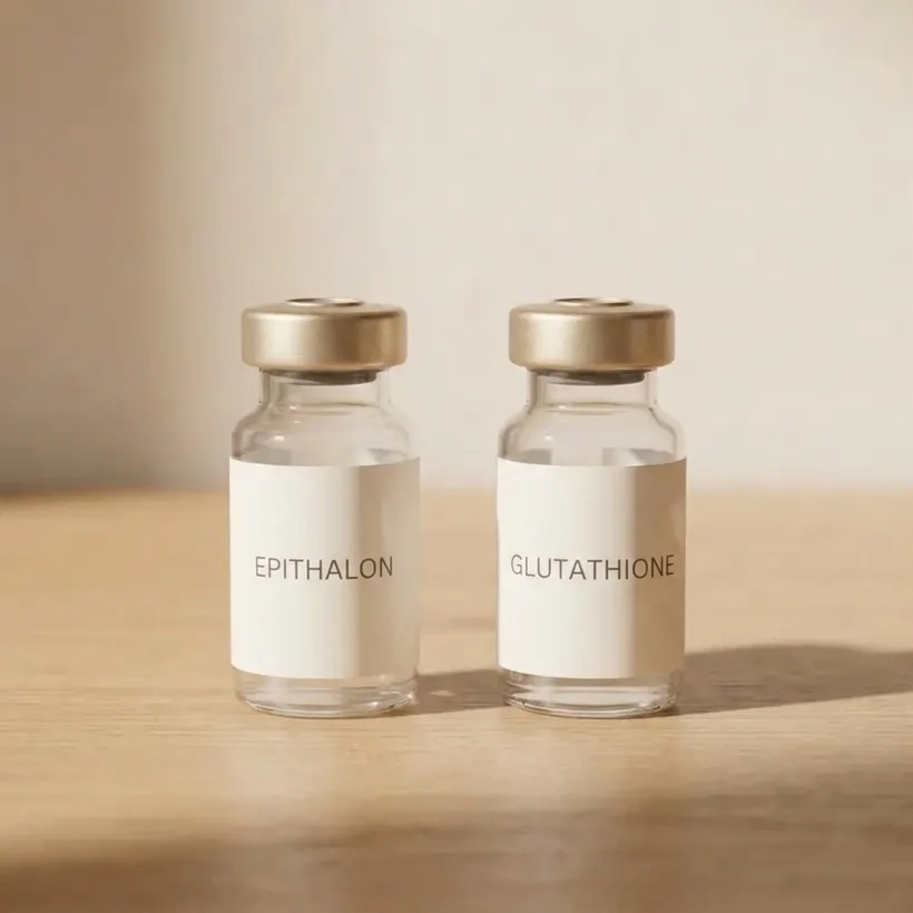
Quick Answer: Anti-aging peptides work by restoring specific signaling pathways that decline with age. They aren't hormones and they aren't supplements. They're bioactive molecules that bind to cellular receptors and trigger defined downstream effects: growth hormone release, collagen gene activation, immune cell maturation, mitochondrial enzyme upregulation, and anti-inflammatory cascading. The science is rooted in endocrinology, immunology, and molecular biology, with evidence ranging from strong (thymosin alpha-1, CJC-1295) to experimental (epithalon, SS-31).
The Science of Peptides and Biological Aging
What Makes a Peptide Different From Other Molecules
A peptide is a chain of amino acids linked by peptide bonds. What distinguishes therapeutic peptides from dietary protein is their specific sequence, which determines their three-dimensional shape, which determines which receptors they bind, which determines their biological effect. The tripeptide GHK (glycine-histidine-lysine) has radically different effects than three random amino acids precisely because its structure allows it to interact with specific cellular targets.
Peptides are naturally occurring in the body. Your pituitary gland produces growth hormone-releasing hormone (GHRH), a 44-amino-acid peptide. Your thymus produces thymosin alpha-1, a 28-amino-acid peptide. Your stomach produces BPC (body protection compound). Therapeutic peptide protocols use synthetic versions of these endogenous molecules (or close analogs) to restore signaling that has declined with age.
The key pharmacological difference between peptides and small-molecule drugs is specificity. A statin blocks HMG-how to read a COA reductase indiscriminately. A peptide like ipamorelin binds selectively to growth hormone secretagogue receptors (GHS-R) and triggers GH release without significantly affecting cortisol, prolactin, or other hormones. This selectivity is the basis for their favorable side effect profiles relative to many pharmaceuticals.
The Hallmarks of Aging: A Scientific Framework
In 2013, Carlos Lopez-Otin and colleagues published a landmark paper in Cell identifying nine hallmarks of aging. Updated in 2023 to twelve hallmarks, this framework provides the scientific basis for understanding why we age and where interventions can be targeted. The hallmarks include:.
- Genomic instability
- Telomere attrition
- Epigenetic alterations
- Loss of proteostasis
- Disabled macroautophagy
- Deregulated nutrient sensing
- Mitochondrial dysfunction
- Cellular senescence
- Stem cell exhaustion
- Altered intercellular communication
- Chronic inflammation
- Dysbiosis
Anti-aging peptides don't address all twelve hallmarks, but they address several with mechanistic precision. Understanding which hallmark each peptide targets helps explain why multi-peptide protocols are more effective than single-peptide approaches and why the choice of peptides should be individualized based on which aging mechanisms are most active in a given person.
The Molecular Mechanisms of Key Anti-Aging Peptides
CJC-1295 and Ipamorelin: The Growth Hormone Axis
The biology of GH decline. Growth hormone is released by somatotroph cells in the anterior pituitary in a pulsatile pattern, with the largest pulse occurring during slow-wave (deep) sleep. Two hypothalamic peptides regulate this release: GHRH stimulates it, and somatostatin inhibits it. With age, GHRH signaling weakens, somatostatin tone increases, and the amplitude of GH pulses decreases by approximately 14% per decade after age 30.
View data table
| Category | Evidence and Efficacy Score | Detail |
|---|---|---|
| Cold Exposure | 72 | Metabolic activation |
| Red Light | 65 | Mitochondrial support |
| CGM Tracking | 82 | Glucose optimization |
| Peptide Stacks | 70 | Targeted protocols |
| Nootropics | 55 | Cognitive enhancement |

This decline, called somatopause, isn't merely cosmetic. GH acts on the liver to stimulate production of IGF-1, which mediates most of GH's peripheral effects: protein synthesis in muscle, lipolysis in adipose tissue, bone mineral deposition, collagen synthesis in skin, and cellular repair processes throughout the body.
How CJC-1295 works. CJC-1295 is a synthetic analog of GHRH (specifically, a modified fragment of GHRH amino acids 1-29) with a Drug Affinity Complex (DAC) that extends its half-life from minutes to approximately 8 days. It binds to GHRH receptors on pituitary somatotrophs and stimulates GH synthesis and release. Because it works through the natural GHRH receptor, it preserves the pulsatile pattern of GH release rather than creating the continuous elevation seen with exogenous GH injection.
Clinical data shows CJC-1295 increases mean GH levels by 2-10 fold and IGF-1 levels by 65-100% after multiple doses, with effects sustained over the dosing interval.
How ipamorelin works. Ipamorelin is a pentapeptide that binds to growth hormone secretagogue receptors (GHS-R), the same receptors that ghrelin (the hunger hormone) activates. But unlike ghrelin, ipamorelin is highly selective for GH release. It doesn't significantly increase cortisol, ACTH, or prolactin at therapeutic doses. This selectivity distinguishes it from earlier GH secretagogues like GHRP-6 and GHRP-2, which had broader hormonal effects.
combined effect. CJC-1295 and ipamorelin work through different receptors (GHRH-R and GHS-R respectively) that converge on the same endpoint: GH release from the pituitary. Using both simultaneously produces a combined effect that exceeds what either peptide achieves alone. This is analogous to pressing the accelerator (CJC-1295) while releasing the brake (ipamorelin reducing somatostatin tone).
Hallmarks addressed: Deregulated nutrient sensing (via IGF-1/GH axis normalization), altered intercellular communication (restoring hormonal signaling), and indirectly, stem cell exhaustion (GH/IGF-1 supports tissue regeneration).
BPC-157: Tissue Repair and the Gut-Systemic Axis
Origin and structure. BPC-157 is a pentadecapeptide (15 amino acids) derived from a protein found in human gastric juice. It's stable in gastric acid, which is unusual for peptides and enables oral administration for gut-specific effects.
Molecular mechanisms. BPC-157's effects are mediated through multiple pathways:
- Nitric oxide system modulation: BPC-157 interacts with the NO system in a context-dependent manner, promoting NO production where healing requires vasodilation and blood flow, and modulating excess NO where inflammation is driving tissue damage.
- Growth factor receptor upregulation: BPC-157 increases expression of VEGF (vascular endothelial growth factor), EGF (epidermal growth factor), and their receptors, promoting angiogenesis and tissue regeneration.
- FAK-paxillin pathway: BPC-157 activates focal adhesion kinase signaling, which is critical for cell migration during wound healing.
- JAK-2/STAT-3 pathway modulation: This pathway is involved in cellular growth, differentiation, and immune regulation. BPC-157's interaction with it contributes to its anti-inflammatory effects.
- Dopamine system interaction: BPC-157 has demonstrated effects on dopamine receptor expression and turnover, which may explain some of its neuroprotective properties observed in animal studies.
The gut-aging connection. One of BPC-157's most relevant anti-aging applications is gut barrier support. Intestinal permeability (leaky gut) increases with age due to tight junction protein degradation, reduced mucosal blood flow, and microbiome shifts. A compromised gut barrier allows bacterial endotoxins (LPS) to enter the bloodstream, driving the chronic low-grade inflammation called inflammaging. BPC-157 directly supports gut mucosal integrity, which makes it an upstream intervention for systemic inflammation.
Evidence base. Over 100 published studies, primarily in animal models. Demonstrated efficacy in healing tendons, ligaments, muscles, gut mucosa, bone, and neural tissue. Human clinical data is limited but clinical use is widespread in longevity and sports medicine practices.
Hallmarks addressed: Chronic inflammation, altered intercellular communication, dysbiosis (via gut barrier support).
Thymosin Alpha-1: Immune Rejuvenation
Thymic biology. The thymus gland is the training ground for T-cells, the adaptive immune system's primary effectors. Thymic tissue is gradually replaced by adipose tissue starting in puberty, a process called thymic involution. By age 50, thymic output of naive T-cells has declined dramatically. This reduces immune surveillance, impairs responses to novel pathogens and vaccines, and reduces the clearance of senescent cells.
How thymosin alpha-1 works. Thymosin alpha-1 (Ta1) is a 28-amino-acid peptide originally isolated from thymic tissue by Allan Goldstein at George Washington University in the 1970s. Its mechanisms include:
- Toll-like receptor activation: Ta1 activates TLR2, TLR5, and TLR9 on dendritic cells and macrophages, enhancing antigen presentation and innate immune activation.
- T-cell maturation: Promotes differentiation of immature T-cells into functional CD4+ and CD8+ populations.
- NK cell activation: Increases natural killer cell cytotoxicity, which is relevant for both anti-viral defense and senescent cell clearance.
- Immune balance: Modulates the Th1/Th2 balance, which is often skewed with age. It doesn't simply stimulate the immune system. it optimizes its function, which is an important distinction for patients with autoimmune tendencies.
Evidence base. FDA orphan drug status. Approved in over 35 countries (as Zadaxin) for hepatitis B and C. Multiple clinical trials demonstrate improved immune response in immunocompromised patients, enhanced vaccine efficacy in elderly populations, and improved outcomes in sepsis. This is one of the most well-characterized peptides in clinical medicine.
Hallmarks addressed: Cellular senescence (via immune surveillance), chronic inflammation (via immune modulation), altered intercellular communication.
GHK-Cu: Gene Expression Reset
Structure and copper binding. GHK-Cu is a naturally occurring tripeptide (glycine-histidine-lysine) that binds copper with high affinity. It was first identified in human plasma by Loren Pickart in 1973, who observed that liver cells exposed to plasma from young individuals grew more vigorously than those exposed to plasma from older individuals. The active factor was isolated and identified as GHK-Cu.
Gene expression modulation. The most striking finding about GHK-Cu comes from gene array studies. Research published in the Journal of Investigative Dermatology and related journals demonstrated that GHK-Cu affects the expression of over 4,000 genes, resetting many of them toward a pattern characteristic of younger tissue. Specifically, it upregulates genes involved in collagen synthesis, DNA repair, and antioxidant defense while downregulating genes associated with inflammation and tissue destruction.
Specific molecular effects:
- Stimulates collagen I, III, and V synthesis
- Increases elastin production
- Promotes glycosaminoglycan (GAG) synthesis, including decorin and dermatan sulfate
- Activates metalloproteinases for controlled tissue remodeling (breaking down damaged collagen to rebuild healthy collagen)
- Increases VEGF for angiogenesis
- Upregulates SOD (superoxide dismutase) and other antioxidant enzymes
- Suppresses TGF-beta driven fibrosis, promoting regeneration over scarring
Evidence base. Extensive in vitro data. Multiple human studies for topical skin applications showing improved skin thickness, elasticity, firmness, and wrinkle reduction. Systemic effects are supported by in vitro and animal data but lack strong human clinical trials.
Hallmarks addressed: Epigenetic alterations (via gene expression modulation), loss of proteostasis (supporting protein turnover in extracellular matrix), altered intercellular communication.
MOTS-c: The Mitochondrial Peptide
Origin. MOTS-c (mitochondrial open reading frame of the 12S rRNA type-c) is unique among therapeutic peptides because it's encoded by mitochondrial DNA, not nuclear DNA. It was discovered in 2015 by Changhan David Lee's group at USC and published in Cell Metabolism.
Mechanism. MOTS-c activates AMPK (AMP-activated protein kinase), the master cellular energy sensor. AMPK activation triggers:
- Increased glucose uptake in skeletal muscle (independent of insulin)
- Enhanced fatty acid oxidation
- Mitochondrial biogenesis via PGC-1alpha activation
- Autophagy activation (cellular cleanup of damaged components)
- Inhibition of mTOR-driven cellular growth (which when chronically activated, accelerates aging)
MOTS-c importantly mimics the metabolic effects of exercise at the molecular level. This doesn't mean it replaces exercise, but it may enhance the metabolic response to exercise and provide metabolic benefits in situations where exercise capacity is limited.
Age-related decline. Circulating MOTS-c levels decrease significantly with age. Studies show reduced MOTS-c in skeletal muscle and plasma of older individuals compared to younger controls. This decline correlates with reduced metabolic flexibility, increased insulin resistance, and impaired exercise capacity.
Evidence base. Strong mechanistic and animal data published in high-impact journals. Human clinical trials are ongoing. The evidence is promising but not yet at the level of CJC-1295 or thymosin alpha-1.
Hallmarks addressed: Mitochondrial dysfunction, deregulated nutrient sensing, disabled macroautophagy.
What to Monitor
- GH axis peptides (CJC-1295/ipamorelin): IGF-1 (primary efficacy marker), IGFBP-3, fasting insulin and glucose (safety monitoring for GH-related insulin resistance)
- Anti-inflammatory peptides (BPC-157): hsCRP, subjective GI symptoms, injury recovery timelines
- Immune peptides (thymosin alpha-1): CBC with differential (lymphocyte subsets), illness frequency, vaccine response if applicable
- Collagen peptides (GHK-Cu): Skin quality assessments (photography under consistent conditions), wound healing speed if relevant
- Mitochondrial peptides (MOTS-c): Fasting insulin, glucose, exercise performance metrics, metabolic flexibility (if RQ testing is available)
- thorough panel every 6 months: Full metabolic, inflammatory, hormonal, and immune markers plus DEXA for body composition
Safety Considerations
- Evidence hierarchy matters. Not all peptides have equal evidence. Thymosin alpha-1 has decades of human clinical data. MOTS-c has strong mechanistic data but limited human trials. Epithalon has interesting animal data but minimal human evidence. Your risk tolerance should scale with the evidence level. Proven peptides first, experimental peptides with informed consent.
- Peptide interactions are possible. Using GH secretagogues alongside peptides that affect insulin sensitivity (like MOTS-c) requires careful metabolic monitoring. GH and AMPK activation have opposing effects on certain metabolic pathways. Your physician needs the full picture.
- Molecular specificity doesn't mean zero side effects. Peptides are more targeted than many drugs, but they still interact with complex biological systems. Individual variation in receptor expression, metabolism, and downstream signaling means that responses will differ between people.
- Don't extrapolate animal data to humans uncritically. Many peptides show dramatic effects in animal models that may or may not translate to humans at the same magnitude. Animal studies are necessary and valuable for understanding mechanisms, but they aren't proof of human efficacy. Be appropriately calibrated in your expectations.
- The dose-response curve isn't linear. More peptide doesn't always mean more effect. Many peptide receptors desensitize with chronic overstimulation. Physiological dosing (mimicking natural levels) often outperforms supraphysiological dosing for the risk-benefit ratio.
Frequently Asked Questions
Do anti-aging peptides actually slow aging?
They address specific mechanisms of aging with measurable biological effects: restoring GH pulsatility, reducing chronic inflammation, supporting immune surveillance, improving mitochondrial function, and stimulating collagen synthesis. Whether this translates to a measurable increase in human lifespan isn't yet proven in clinical trials. What is proven is that these interventions improve biomarkers associated with aging and improve functional outcomes (body composition, immune function, recovery, metabolic health) that deteriorate with age.
From the FormBlends catalog
Longevity Reset
Telomere support with the master antioxidant. · Bundle from $270 · provider-reviewed peptide bundle.
View Longevity Reset →Why not just take exogenous growth hormone?
Exogenous GH provides continuous, non-pulsatile elevation that suppresses your pituitary's natural release through negative feedback. It also carries higher risks of insulin resistance, fluid retention, joint pain, and potentially cancer at supraphysiological doses. GH secretagogue peptides stimulate your own pituitary to release GH in its natural pulsatile pattern, maintaining feedback regulation. The safety profile is more favorable and the physiological pattern is more natural. Exogenous GH has legitimate medical uses for true GH deficiency, but for improvement in aging, secretagogues are generally preferred.
Can peptides replace exercise for anti-aging?
No. Exercise produces a breadth of anti-aging effects that no peptide or combination of peptides can replicate: mitochondrial biogenesis, neuroplasticity (via BDNF), cardiovascular adaptation, glucose disposal, bone loading, hormonal improvement, and psychological resilience. MOTS-c mimics some metabolic effects of exercise at the molecular level, but it can't reproduce the mechanical, cardiovascular, or neurological benefits. Peptides and exercise are combined, not substitutes.
How do scientists measure whether an anti-aging intervention is working?
Several approaches exist. Biological age clocks (epigenetic clocks like GrimAge and DunedinPACE) estimate biological age based on DNA methylation patterns. Biomarker panels track physiological function (insulin sensitivity, inflammatory markers, immune cell populations). Functional assessments measure grip strength, VO2 max, cognitive performance, and other age-sensitive metrics. The gold standard would be a randomized controlled trial measuring lifespan, but human longevity studies take decades. In practice, interventions are evaluated by their effects on validated surrogates of aging.
What is the difference between anti-aging peptides and senolytics?
Senolytics (like dasatinib + quercetin or fisetin) target cellular senescence by selectively killing senescent cells that accumulate with age and secrete inflammatory factors (the SASP, or senescence-associated secretory phenotype). Anti-aging peptides generally work by restoring signaling and function rather than eliminating cells. Thymosin alpha-1 bridges these categories by enhancing immune surveillance that clears senescent cells. The two approaches are complementary rather than competing, addressing different hallmarks of aging.
Put the Science to Work
About the molecular mechanisms behind anti-aging peptides isn't academic exercise. It's the basis for rational protocol design. At FormBlends, our physician-supervised telehealth platform uses this science to design peptide protocols matched to your biology, your biomarkers, and your aging trajectory. Every protocol is built on evidence and monitored with data.
Begin your consultation at FormBlends.com and access science-driven anti-aging medicine.
See your options in about 2 minutes
Take the free quiz and see what fits you. Quick, private, and no commitment to continue.
See my options →