Executive Summary
Key Takeaways
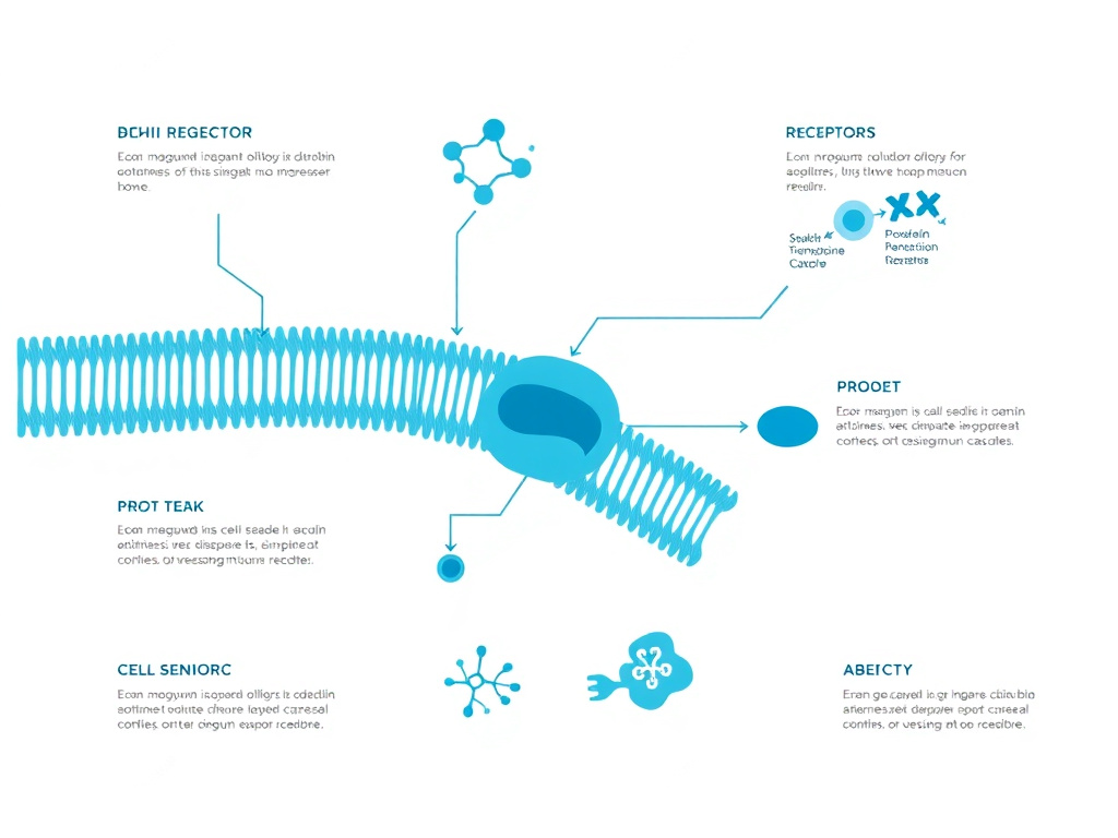
- Figure 1: Epithalon (AEDG) is a synthetic tetrapeptide derived from the pineal gland extract epithalamin, designed to activate telomerase and modulate aging pathways.
- Epithalon (also spelled epitalon or epithalone) is a synthetic tetrapeptide with the amino acid sequence Ala-Glu-Asp-Gly (AEDG).
- Developed by Professor Vladimir Khavinson at the St.
- Petersburg Institute of Bioregulation and Gerontology, it represents one of the most extensively studied peptide bioregulators in the anti-aging research space.
- Its primary mechanism centers on the activation of telomerase, the enzyme responsible for maintaining telomere length at the ends of chromosomes, which directly influences cellular replicative capacity and biological aging.
Epithalon (also spelled epitalon or epithalone) is a synthetic tetrapeptide with the amino acid sequence Ala-Glu-Asp-Gly (AEDG). Developed by Professor Vladimir Khavinson at the St. Petersburg Institute of Bioregulation and Gerontology, it represents one of the most extensively studied peptide bioregulators in the anti-aging research space. Its primary mechanism centers on the activation of telomerase, the enzyme responsible for maintaining telomere length at the ends of chromosomes, which directly influences cellular replicative capacity and biological aging.
The story of epithalon begins with epithalamin, a polypeptide complex originally extracted from bovine pineal glands. Khavinson and his team at the Russian Institute of Bioregulation and Gerontology spent decades characterizing epithalamin's effects on aging, pineal function, and immune regulation. From that work emerged the identification of AEDG as the minimal bioactive sequence responsible for many of epithalamin's observed benefits. This synthetic tetrapeptide could be produced with greater consistency and purity than bovine-derived extracts, making it a more practical tool for both research and potential clinical application.
What sets epithalon apart from many other anti-aging compounds is the breadth of its proposed biological activity. At the cellular level, it reactivates telomerase expression in somatic cells that have normally silenced the enzyme. In landmark in vitro work, Khavinson's group demonstrated that human fetal fibroblasts treated with epithalon expressed the catalytic subunit of telomerase (hTERT), developed measurable enzymatic activity, and elongated their telomeres by an average of 33.3%. Control cultures lost the ability to divide after passage 34, while epithalon-treated cells continued proliferating past passage 44. These findings, published in the Bulletin of Experimental Biology and Medicine in 2003, positioned epithalon as one of the first peptides shown to directly reactivate telomerase in normal human somatic cells.
Beyond telomere maintenance, epithalon exerts significant effects on the pineal gland and melatonin production. The pineal gland is a neuroendocrine organ that produces melatonin, the hormone governing circadian rhythms, sleep architecture, and a wide range of antioxidant and immunomodulatory functions. As organisms age, pineal function declines and melatonin output drops, a process closely correlated with age-related disease progression. In both animal models and human studies, epithalon and its parent compound epithalamin have been shown to restore melatonin secretion to more youthful levels, normalize circadian cortisol rhythms, and upregulate key antioxidant enzymes including superoxide dismutase and glutathione peroxidase.
Animal longevity data, while generated primarily from a single research group, remain striking. In female Swiss-derived SHR mice, epithalon treatment increased the lifespan of the last 10% of survivors by 13.3% and maximum lifespan by 12.3%. The compound also reduced leukemia incidence by 6-fold and decreased chromosome aberrations in bone marrow cells by 17.1%. Studies in Drosophila and rats have shown similar trends toward extended mean and maximal lifespan. In one of the most ambitious human observational studies, elderly patients treated with epithalamin combined with thymalin (a thymic peptide) over six years experienced a 4.1-fold decrease in mortality compared to controls during a 15-year follow-up period.
The safety profile of epithalon appears favorable based on available data. No significant adverse effects were reported in the 15-year follow-up of elderly patients. Common side effects in current usage are limited to mild injection site reactions, occasional headaches, and transient fatigue. Theoretical concerns about telomerase activation promoting cancer growth have not been confirmed in animal studies, and several investigations have actually demonstrated reduced tumor incidence in epithalon-treated animals.
Standard dosing protocols call for 5-10 mg administered subcutaneously once daily for 10-20 consecutive days, with the cycle repeated once or twice per year. Evening administration is often preferred to align with natural melatonin production rhythms. This report examines the full scope of epithalon research, covering its history, molecular mechanisms, preclinical and clinical evidence, practical dosing guidance, and safety considerations. For those interested in related longevity compounds, the peptide research hub provides additional context on complementary approaches to biological aging.
A critical caveat must be stated at the outset: the vast majority of epithalon research originates from Professor Khavinson's group in Russia. While the body of work is extensive, spanning hundreds of publications across several decades, independent replication by outside laboratories has been limited. A 2025 study published in Biogerontology by researchers at the Leibniz Institute provided some of the first independent confirmation of epithalon's telomere-lengthening effects in human cell lines, representing an important step toward broader validation. Readers and clinicians should weigh the existing evidence with this context in mind.
Positioning Epithalon Within the Hallmarks of Aging Framework
Modern aging research is organized around the "hallmarks of aging" framework, first proposed by Lopez-Otin and colleagues in 2013 and updated in 2023 with additional hallmarks. These hallmarks represent the fundamental biological processes that drive aging across organisms, and they include genomic instability, telomere attrition, epigenetic alterations, loss of proteostasis, deregulated nutrient sensing, mitochondrial dysfunction, cellular senescence, stem cell exhaustion, altered intercellular communication, disabled macroautophagy, chronic inflammation, and dysbiosis.
Epithalon's appeal lies in its potential to address multiple hallmarks simultaneously. Its telomerase activation directly targets telomere attrition, one of the primary hallmarks. By preventing or delaying cellular senescence (a downstream consequence of telomere shortening), it indirectly addresses the cellular senescence hallmark. Its effects on gene expression and chromatin regulation may intersect with epigenetic alterations. And its restoration of melatonin production and neuroendocrine function touches on altered intercellular communication and the emerging hallmark of chronic inflammation, given melatonin's potent anti-inflammatory properties.
Few individual compounds address more than one or two hallmarks of aging. The multi-target profile attributed to epithalon, if validated by further research, would place it among a select group of interventions with broad anti-aging potential. Other compounds in this category include NAD+ precursors (which target mitochondrial dysfunction, DNA repair, and epigenetic regulation), rapamycin (which targets deregulated nutrient sensing through mTOR inhibition), and metformin (which affects nutrient sensing, inflammation, and cellular senescence through AMPK activation). The challenge with each of these compounds, epithalon included, is translating promising mechanistic data into clinically validated human outcomes.
The research community's growing recognition that aging is a treatable condition rather than an inevitable decline has created a more receptive environment for compounds like epithalon. As clinical aging biomarkers become more validated and regulatory agencies begin to consider aging as a legitimate therapeutic target, the pathway from research compound to approved therapy may become more navigable. For now, epithalon occupies an interesting space: extensively researched within a specific academic tradition, mechanistically plausible, supported by suggestive animal and human data, but still awaiting the large-scale controlled trials that would establish its efficacy by Western medical standards.
Who Uses Epithalon Today
Current epithalon users span several demographics. Longevity-focused clinicians, sometimes operating within the functional medicine or anti-aging medicine space, prescribe epithalon as part of comprehensive longevity protocols for patients interested in proactive health optimization. Biohackers and self-experimenters obtain the peptide through research chemical suppliers and track their own biomarkers before and after treatment cycles. In Russia and some Eastern European countries, epithalamin and related peptide bioregulators are used in mainstream clinical practice for geriatric patients. And a growing number of integrative medicine practitioners worldwide are incorporating epithalon into treatment plans for patients with specific concerns about aging, sleep quality, or immune function.
The regulatory status of epithalon varies by country and is evolving. In Russia, the parent compound epithalamin has been an approved pharmaceutical for decades. In most Western countries, epithalon is available as a research compound or through compounding pharmacies under physician supervision. In the United States, the FDA's evolving guidance on compounded peptides has created some uncertainty about the long-term availability of certain peptides, though epithalon has not been specifically targeted by regulatory action as of early 2026. Clinicians prescribing peptide therapies should stay informed about regulatory developments in their jurisdiction. The GLP-1 overview page provides context on how the regulatory environment for peptide therapies is developing.
The typical epithalon user is health-conscious, often already engaged in other longevity practices (exercise, nutrition optimization, supplementation), and willing to accept a degree of scientific uncertainty in exchange for potential anti-aging benefits. This profile is similar to early adopters of other peptide therapies, including BPC-157 for tissue healing and CJC-1295/Ipamorelin for growth hormone optimization. The free assessment can help determine whether peptide therapy might be appropriate for your individual health goals.
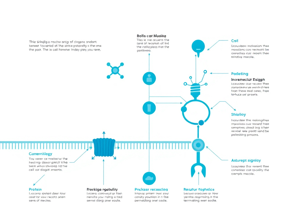
Khavinson's Peptide Research
Figure 2: The evolution of peptide bioregulation research under Professor Vladimir Khavinson spans over five decades of systematic investigation.
Origins of Peptide Bioregulation Theory
Vladimir Khavinson's career in peptide bioregulation stretches back to the early 1970s, when he began investigating the role of short peptides in organ-specific regulation at the Military Medical Academy in Leningrad (now St. Petersburg). The conceptual foundation rested on a straightforward observation: organs produce small peptides that regulate their own function and communicate with other organ systems. When organs age or become dysfunctional, their peptide output declines, contributing to systemic deterioration. Restoring these peptides, either through administration of organ extracts or synthetic analogs, might reverse or slow age-related decline.
This framework led Khavinson to isolate and characterize dozens of peptide preparations from various animal organs. The earliest and most prominent were thymalin, derived from the thymus gland, and epithalamin, derived from the pineal gland. Each organ extract contained a complex mixture of short peptides, proteins, and other bioactive molecules. The challenge was determining which specific sequences drove the observed biological effects.
Khavinson's early military research focused on thymalin as an immune-boosting agent. The Soviet military was keenly interested in radiation protection and immune recovery, making thymic peptides a strategic priority. The success of thymalin in clinical trials through the 1980s established Khavinson's credibility and provided the institutional support needed to expand his research into pineal peptides and aging.
Discovery and Characterization of Epithalamin
Epithalamin emerged from Khavinson's systematic extraction of bovine pineal glands in the late 1970s and early 1980s. The crude extract demonstrated a remarkable range of biological effects: it increased melatonin production, normalized circadian rhythms, enhanced antioxidant defense systems, and appeared to extend lifespan in animal models. But epithalamin was a complex mixture, difficult to standardize, and potentially immunogenic due to its animal-derived components.
Through progressive purification and analysis, Khavinson's team identified the tetrapeptide sequence Ala-Glu-Asp-Gly as a key bioactive component of epithalamin. This synthetic version, initially designated Epitalon (and later also known as epithalon or epithalone), could be produced with consistent purity and potency. Early comparative studies suggested that the synthetic tetrapeptide reproduced many of epithalamin's effects, including its influence on melatonin production, telomerase activity, and aging biomarkers.
The transition from natural extract to synthetic peptide was significant for several reasons. First, it eliminated batch-to-batch variability inherent in animal-derived preparations. Second, it reduced the risk of prion contamination or allergic reactions associated with bovine tissue products. Third, it made large-scale production feasible. And fourth, it allowed researchers to study a single, defined molecular entity rather than a complex mixture, strengthening the ability to attribute observed effects to a specific compound.
The St. Petersburg Institute of Bioregulation and Gerontology
In 1992, Khavinson founded the St. Petersburg Institute of Bioregulation and Gerontology, which became the primary center for peptide bioregulation research worldwide. The institute has published hundreds of papers on short peptides, covering everything from cellular mechanisms to large-scale clinical observations. Khavinson's publication record is extraordinary in scope, with over 800 scientific papers and numerous monographs to his name.
The institute developed a systematic framework for what Khavinson terms "peptide bioregulation." The core theory posits that short peptides (2-7 amino acids in length) can penetrate cell nuclei, interact with DNA at specific promoter regions, and regulate gene expression in an organ-specific manner. This concept goes beyond simple receptor-mediated signaling. Khavinson's group has demonstrated through molecular modeling and in vitro studies that short peptides can form complexes with DNA double helices, potentially influencing transcription in a sequence-dependent fashion.
The institute's research program is not limited to epithalon. Dozens of organ-specific peptide bioregulators have been characterized, including pinealon (for brain function), vesugen (for vascular health), cartalax (for cartilage), and many others. Each follows the same development pipeline: extraction from target organ tissue, identification of the minimal active peptide sequence, chemical synthesis, and biological characterization. This systematic approach has generated a large body of data, though it has also concentrated nearly all the evidence within a single research institution.
Regulatory Status and International Recognition
In Russia, epithalamin and thymalin received regulatory approval as pharmaceutical agents decades ago. They have been used clinically in thousands of patients, primarily for immune support and anti-aging applications. Khavinson has received significant state recognition for his work, including the Laureate of the President of Russia Prize and membership in the Russian Academy of Sciences.
Outside Russia, however, epithalon remains an unregulated research compound. It has not been submitted for FDA approval in the United States, nor has it undergone the randomized controlled trials required by Western regulatory agencies. The compound is available through compounding pharmacies and research chemical suppliers, but its legal status varies by jurisdiction. In the United States, it can be obtained for research purposes or through licensed compounding pharmacies with a prescription. Those considering epithalon should work with a qualified healthcare provider familiar with peptide therapies.
International interest in Khavinson's work has grown substantially since the 2010s, driven partly by the broader longevity research movement and the increasing availability of information about peptide bioregulators online. Academic citations of his work have increased, and several review articles in Western journals have provided critical assessments of the evidence base. The 2025 independent replication study by German researchers represented a milestone in moving beyond reliance on a single laboratory's findings.
Legacy and Continuing Research
As of 2026, Khavinson's research program continues actively. Recent publications have explored epithalon's effects on gene expression patterns, particularly circadian rhythm genes such as Clock, Csnk1e, and Cry2 in human leukocytes and blood lymphocytes. Additional work has examined the peptide's interactions with DNA at the molecular level using computational modeling and biophysical techniques.
The broader field of peptide bioregulation remains somewhat isolated from mainstream Western pharmacology. The language barrier (many early publications were in Russian), the concentration of research within a single institute, and the lack of pharmaceutical industry interest (short peptides are difficult to patent) have all contributed to limited international adoption. Yet the growing availability of peptides through compounding pharmacies and research suppliers has created a grassroots interest among longevity-focused clinicians and biohackers. For those exploring the broader world of anti-aging peptide research, the biohacking hub provides additional resources on related compounds and protocols.
Understanding Khavinson's contribution requires appreciating both the ambition and the limitations of his work. The peptide bioregulation framework is comprehensive, internally consistent, and supported by an enormous volume of publications. But the academic world rightly demands independent replication, and epithalon's journey from a Russian laboratory curiosity to a globally recognized anti-aging compound depends on ongoing validation by independent research groups.
The Broader Peptide Bioregulator Family
Epithalon exists within a larger family of short peptide bioregulators developed by Khavinson's institute. Understanding this family provides context for epithalon's development and its place in the peptide bioregulation framework. Each peptide bioregulator is organ-specific, meaning it was derived from and acts upon a particular organ or tissue system. The full catalog includes dozens of compounds, though only a handful have received extensive investigation.
Thymalin (EW dipeptide) targets the thymus and immune system. It was the first peptide bioregulator to receive widespread clinical use, beginning in the 1970s for immune deficiency states, radiation exposure, and post-surgical immune recovery. In Khavinson's longevity studies, thymalin was frequently used in combination with epithalon, and the paired treatment consistently produced better outcomes than either compound alone. This additive benefit suggests that addressing both immune and pineal decline simultaneously may be more effective than targeting either system in isolation.
Pinealon (EDR tripeptide) is a more recently characterized pineal-targeted peptide that shows neuroprotective properties. Unlike epithalon, which primarily affects telomerase and melatonin production, pinealon has been studied for its effects on cognitive function, neuroprotection, and stress resilience. The two pineal peptides may work through overlapping but distinct pathways, with epithalon focused on longevity-related mechanisms and pinealon on neurological function. Related neuroprotective peptides available for research include Semax, Selank, and Dihexa.
Vesugen (KED tripeptide) was identified as a vascular-targeted bioregulator with effects on endothelial function, blood vessel integrity, and cardiovascular health. Cartalax (AED tripeptide) targets cartilage and musculoskeletal tissue. Pancragen (KEDW tetrapeptide) is directed at pancreatic function and blood sugar regulation. Crystagen (EDP tripeptide) supports immune function through pathways distinct from thymalin. Each of these bioregulators follows the same development paradigm: extraction from the target organ, identification of the active peptide sequence, chemical synthesis, and biological characterization.
The peptide bioregulator family illustrates a distinctive approach to drug development that differs markedly from the Western pharmaceutical model. Rather than screening large chemical libraries for molecules that hit a single molecular target, Khavinson's approach starts with the biology of organ-specific regulation and identifies the endogenous peptide signals that maintain healthy function. The result is a pharmacopoeia of short peptides, each with broad, organ-specific regulatory effects rather than precise single-target activity. This approach has both strengths (potentially addressing multiple age-related changes within an organ system) and weaknesses (difficulty attributing effects to specific molecular mechanisms, challenges with clinical trial design).
DNA-Peptide Interaction Studies
A distinctive aspect of Khavinson's research program has been the investigation of how short peptides interact directly with DNA. This work has proceeded through a combination of experimental techniques (fluorescence labeling, gel shift assays, spectrophotometry) and computational methods (molecular docking, molecular dynamics simulations). The findings have been published in several high-profile journals including Molecules, Russian Journal of Genetics, and Biochemistry (Moscow).
Using fluorescence-labeled peptides, Khavinson's group demonstrated that short peptides (2-4 amino acids) can penetrate into cell nuclei and nucleoli of cultured human cells. The peptides were observed to associate with chromatin and to localize in specific nuclear compartments. This nuclear penetration is plausible given the small size of these peptides: at 390 daltons, AEDG is well below the approximately 40,000 dalton cutoff for passive diffusion through nuclear pores, and its small size also facilitates passive membrane crossing.
In vitro binding studies showed that AEDG and other short peptides form stable complexes with DNA sequences in gene promoter regions. The binding appeared to show some sequence preference, with the negatively charged residues (glutamate and aspartate) in the peptide interacting with positively charged regions of the DNA minor groove. The binding affinities were in the micromolar range, which is orders of magnitude weaker than the nanomolar affinities typical of transcription factor-DNA interactions but potentially relevant at the high local concentrations that might be achieved inside cell nuclei.
A systematic review published in Molecules in 2021 compiled the evidence for peptide regulation of gene expression across dozens of studies from multiple research groups. The review concluded that short peptides can influence gene expression through both direct DNA interactions and indirect mechanisms involving membrane receptors, intracellular signaling cascades, and epigenetic modifications. The multi-modal mechanism of action may explain why the same peptide can produce effects on diverse targets including telomerase, melatonin synthesis enzymes, and circadian clock genes.
Critical Assessment of the Evidence Base
Any honest evaluation of epithalon must grapple with the unusual concentration of evidence within a single research group. In most fields of biomedical research, a finding is considered established only after independent replication by multiple laboratories. The epithalon literature departs from this norm in significant ways.
First, virtually all animal longevity studies come from Khavinson's group and close collaborators, primarily at the N.N. Petrov Research Institute of Oncology (where Anisimov led the cancer-related studies). No independent laboratory has published a lifespan study with epithalon in any species.
Second, the human clinical data come from observational studies with significant methodological limitations. The 15-year longevity study was not randomized, not blinded, and not controlled in the manner required by contemporary clinical trial standards. Patient selection, outcome assessment, and statistical analysis may have been subject to biases that are difficult to assess from the published reports.
Third, many of the original publications were in Russian-language journals or in journals with limited international peer review. While this doesn't necessarily indicate poor quality, it does mean that the work has received less external scrutiny than studies published in major English-language journals.
Fourth, there is a potential for publication bias. Khavinson's group has published hundreds of papers on peptide bioregulators, overwhelmingly reporting positive results. The possibility that negative or null results were not published cannot be excluded. This is a concern common to many areas of biomedical research, but it is particularly relevant when the evidence base is dominated by a single group.
Against these concerns, several factors support the credibility of the evidence. The body of work is internally consistent across decades and multiple experimental systems. The proposed mechanisms are biologically plausible and align with established understanding of telomere biology, pineal function, and aging. The 2025 independent replication study provided objective confirmation of at least one key claim. And the safety data from long-term use in elderly patients, while observational, provide meaningful real-world evidence. For researchers and clinicians, the appropriate stance is cautious optimism: the data are promising enough to warrant continued investigation but not sufficient to make definitive clinical claims.
Telomere Biology
What Are Telomeres?
Telomeres are specialized nucleoprotein structures located at the ends of linear chromosomes. In vertebrates, they consist of thousands of tandem repeats of the hexanucleotide sequence 5'-TTAGGG-3', bound by a multi-protein complex called shelterin. Together, the DNA repeats and their associated proteins form a protective cap that prevents chromosome ends from being recognized as double-strand DNA breaks, which would trigger DNA damage repair pathways, chromosomal fusions, and genomic instability.
The structure of telomeric DNA is more complex than a simple linear extension. The 3' end of the telomere forms a single-stranded overhang of approximately 150-200 nucleotides, known as the G-overhang. This overhang can invade the double-stranded telomeric region, forming a large loop structure called a T-loop. The displaced strand at the invasion site creates a smaller displacement loop, or D-loop. This looped configuration effectively hides the chromosome end from the cellular machinery that scans for DNA damage.
Human telomeres at birth typically measure 10,000-15,000 base pairs (10-15 kilobases). They shorten with each cell division at a rate of approximately 50-200 base pairs per division, depending on cell type, oxidative stress exposure, and other factors. When telomeres reach a critical minimum length, generally around 4-6 kilobases, the cell enters a state of permanent growth arrest known as replicative senescence. This process forms the molecular basis of the Hayflick limit, the observation that normal human cells have a finite number of divisions before they stop proliferating.
The Shelterin Complex
The shelterin complex consists of six core proteins that associate specifically with telomeric DNA: TRF1, TRF2, POT1, TIN2, TPP1, and RAP1. Each plays a distinct role in telomere maintenance and protection. TRF1 and TRF2 bind directly to double-stranded TTAGGG repeats. POT1 binds the single-stranded G-overhang. TIN2 serves as a bridging protein connecting TRF1, TRF2, and TPP1. TPP1 links POT1 to TIN2 and helps recruit telomerase. RAP1, recruited by TRF2, assists in telomere length regulation.
When shelterin components are depleted or displaced from telomeres, the exposed chromosome ends activate the ATM and ATR DNA damage signaling kinases. This triggers a cascade that can lead to cell cycle arrest, apoptosis, or chromosomal end-to-end fusions. The importance of shelterin is demonstrated by the severe consequences of its disruption: loss of TRF2, for example, results in rapid telomere uncapping, activation of the non-homologous end-joining pathway, and widespread chromosome fusions.
Shelterin also regulates access of telomerase to telomere ends. TRF1, through its effects on telomere structure, acts as a negative regulator of telomere length by limiting telomerase access. TPP1 and POT1, in contrast, can enhance telomerase processivity. This regulatory balance ensures that telomere length is maintained within a functional range in cells that express telomerase, such as stem cells and germ cells.
The End Replication Problem
The progressive shortening of telomeres with each cell division is a direct consequence of a fundamental limitation in DNA replication called the end replication problem. During DNA synthesis, the lagging strand is replicated discontinuously through the formation of short RNA primers (Okazaki fragments). The removal of the terminal RNA primer at the 5' end of the lagging strand leaves a gap that cannot be filled by DNA polymerase, because no upstream sequence exists to provide a 3' hydroxyl group for polymerase extension.
The result is that every round of DNA replication produces daughter chromosomes slightly shorter than their parent. This shortening is not random; it occurs specifically at the chromosome ends. In the absence of a compensatory mechanism, this progressive erosion would eventually eat into essential coding regions of the genome, causing gene loss and cell death.
Telomeres solve this problem by providing a buffer of non-coding, repetitive DNA at chromosome ends. The cell can afford to lose 50-200 base pairs of TTAGGG repeats per division without any functional consequence, because these sequences don't encode genes. But this buffer is finite. After a sufficient number of divisions, the buffer is consumed, and the cell's replicative clock expires. This mechanism functions as a tumor suppressor, limiting the proliferative potential of cells and reducing the risk of accumulated mutations during repeated cell divisions. For a broader look at how cellular aging relates to health, visit the science and research section.
Telomere Length as a Biomarker of Aging
Epidemiological studies have consistently demonstrated an inverse correlation between telomere length and chronological age. Leukocyte telomere length (LTL), measured from peripheral blood white blood cells, serves as the most commonly used clinical metric. Average LTL declines from approximately 11 kilobases at birth to around 7 kilobases by age 70, though there is substantial individual variation.
Shorter telomere length has been associated with increased risk of cardiovascular disease, type 2 diabetes, certain cancers, neurodegenerative diseases, and all-cause mortality. The Cawthon study in 2003, examining 143 individuals over age 60, found that those with shorter telomeres had significantly higher mortality rates from heart disease and infectious diseases. Subsequent large-scale studies, including analyses from the UK Biobank involving hundreds of thousands of participants, have confirmed these associations.
However, the relationship between telomere length and disease is not strictly causal. Telomere shortening is influenced by genetics (approximately 70-80% of telomere length variation is heritable), lifestyle factors (smoking, obesity, sedentary behavior, chronic stress), and disease states themselves. Shorter telomeres may be both a cause and a consequence of poor health, making it difficult to determine directionality. Mendelian randomization studies have provided some evidence for causal relationships in specific diseases, but the picture remains complex.
The measurement of telomere length is itself subject to methodological challenges. Quantitative PCR (qPCR), the most widely used method, provides a ratio of telomere repeat copy number to single-copy gene copy number, rather than an absolute measurement. Terminal restriction fragment (TRF) analysis by Southern blot gives a more direct measurement but requires more DNA and is less scalable. Newer techniques such as STELA (single telomere length analysis) and Flow-FISH offer improved resolution but are technically demanding. Differences in methodology can produce significantly different results, making cross-study comparisons challenging.
Despite these limitations, telomere length has emerged as one of the most studied biomarkers in aging research. The appeal is straightforward: telomere shortening is mechanistically linked to cellular senescence, and interventions that slow or reverse telomere attrition could, in theory, extend both cellular and organismal healthspan. This is the fundamental premise underlying epithalon research and its potential role in longevity science. Individuals interested in how biological aging relates to overall health optimization may find the lifestyle hub a useful complementary resource.
Cellular Senescence and Its Consequences
When telomeres shorten below a critical threshold, cells enter replicative senescence, a state of permanent growth arrest first described by Leonard Hayflick in 1961. Senescent cells remain metabolically active but lose the ability to divide. They also develop a distinctive phenotype characterized by increased cell size, flattened morphology, expression of senescence-associated beta-galactosidase (SA-beta-gal), and upregulation of cell cycle inhibitors p21 and p16INK4a.
Perhaps most significant for systemic aging is the senescence-associated secretory phenotype (SASP). Senescent cells secrete a complex mixture of pro-inflammatory cytokines (IL-6, IL-8, TNF-alpha), matrix metalloproteinases, growth factors, and chemokines. This SASP drives chronic low-grade inflammation, sometimes called "inflammaging," which contributes to tissue dysfunction, fibrosis, and disease progression across multiple organ systems.
The accumulation of senescent cells with age has been convincingly linked to age-related pathology in animal models. Transgenic mice engineered to selectively clear senescent cells (the INK-ATTAC model developed by the Mayo Clinic) showed delayed onset of age-related conditions including cataracts, sarcopenia, and renal dysfunction. This finding sparked intense interest in senolytic therapies, drugs that selectively kill senescent cells, as well as strategies to prevent senescence from occurring in the first place.
Epithalon's proposed mechanism addresses senescence at its root cause. By reactivating telomerase and preventing telomere shortening, the peptide could theoretically delay the onset of replicative senescence, reducing the accumulation of senescent cells and their associated SASP. This approach differs fundamentally from senolytics, which clear senescent cells after they have formed. A preventive strategy that maintains telomere length could, in principle, reduce the overall burden of cellular senescence across a lifetime. Related compounds with anti-aging potential include NAD+ and MOTS-c, which address cellular aging through complementary mechanisms.
Telomere Length Measurement Methods and Their Limitations
Understanding the strengths and weaknesses of telomere measurement methods is essential for evaluating epithalon research claims. The field uses several techniques, each with distinct advantages and limitations that affect how results should be interpreted.
Quantitative PCR (qPCR), the most widely used method in population studies, measures the ratio of telomere repeat copy number (T) to a single-copy gene reference (S), expressed as a T/S ratio. This method is scalable, requires only small amounts of DNA, and can be performed with standard laboratory equipment. However, it provides a relative measurement rather than an absolute telomere length, is sensitive to DNA quality and quantity, and has a coefficient of variation that can exceed 10% between replicates. Results from different laboratories using different qPCR protocols are not always directly comparable.
Terminal Restriction Fragment (TRF) analysis, performed by Southern blotting, provides a more direct measurement of telomere length. DNA is digested with restriction enzymes that cut in subtelomeric regions, and the resulting terminal fragments are separated by gel electrophoresis and detected with a telomere-specific probe. TRF gives a distribution of fragment sizes rather than a single number, reflecting the heterogeneity of telomere lengths across chromosomes and cells. The main limitations are the requirement for larger amounts of DNA, lower throughput, and inclusion of subtelomeric DNA in the measurement, which can inflate apparent telomere length.
Flow-FISH (fluorescence in situ hybridization with flow cytometry) measures telomere fluorescence in individual cells, providing cell-type-specific telomere length data. This technique is particularly useful for immunological studies, as it can distinguish telomere length in different lymphocyte populations. It is the basis of the most clinically validated telomere measurement services. However, it requires fresh blood samples and specialized equipment, limiting its accessibility.
Single Telomere Length Analysis (STELA) measures the length of individual telomeres at specific chromosome ends, providing the most detailed picture of telomere length distribution. This technique can detect critically short telomeres that might be masked in average measurements, making it particularly informative for understanding senescence risk. Its limited throughput makes it primarily a research tool.
When evaluating epithalon's telomere-lengthening claims, the measurement method used matters substantially. The 33.3% increase reported in Khavinson's original study and the dose-dependent lengthening observed in the 2025 replication study used different techniques, and the absolute magnitude of change should be interpreted within the context of each method's precision and accuracy. Future studies using multiple measurement approaches on the same samples would provide the strongest evidence for epithalon's effects on telomere length.
Telomere Biology and Disease Risk: Current Evidence
The relationship between telomere length and disease extends across virtually every major age-related condition. Large-scale epidemiological studies and Mendelian randomization analyses have clarified these associations, providing context for understanding why telomere-lengthening interventions like epithalon generate such interest.
In cardiovascular disease, shorter leukocyte telomere length is associated with increased risk of coronary artery disease, heart failure, and stroke. A meta-analysis of 24 studies involving over 43,000 participants found that individuals in the shortest telomere length tertile had a 40-50% higher risk of coronary heart disease compared to those in the longest tertile. Mendelian randomization studies using genetic variants associated with telomere length have provided evidence for a causal relationship in coronary artery disease, though the effect size is modest.
For type 2 diabetes, shorter telomeres have been associated with insulin resistance, impaired beta-cell function, and incident diabetes. Chronic hyperglycemia accelerates telomere shortening through oxidative stress, creating a potential feedback loop where metabolic dysfunction drives further telomere erosion. For those interested in metabolic health optimization, semaglutide and tirzepatide represent evidence-based approaches to glycemic control that may indirectly support telomere maintenance by reducing metabolic stress.
In neurodegeneration, shorter telomeres have been linked to Alzheimer's disease, Parkinson's disease, and cognitive decline. The brain is particularly vulnerable to the consequences of cellular senescence, as senescent glial cells produce inflammatory mediators that can damage neurons and accelerate neurodegeneration. Whether telomere-lengthening interventions could modify neurodegenerative disease risk remains speculative but represents an active area of investigation.
Cancer has the most complex relationship with telomere biology. Short telomeres increase genomic instability, which can promote cancer initiation. But telomerase activation, which prevents telomere shortening, is also a hallmark of established cancers. This dual relationship means that the optimal telomere maintenance strategy for cancer prevention is unclear and likely depends on the specific cancer type, genetic background, and timing of intervention.
Telomerase Activation Mechanism
The Telomerase Enzyme Complex
Telomerase is a specialized ribonucleoprotein enzyme that adds TTAGGG repeats to the 3' ends of chromosomes, counteracting the end replication problem. The enzyme consists of two essential components: the telomerase reverse transcriptase (TERT, or hTERT in humans), which provides the catalytic activity, and the telomerase RNA component (TERC, or hTERC in humans), which serves as the template for telomeric repeat synthesis. Additional accessory proteins, including dyskerin, NOP10, NHP2, and GAR1, stabilize the complex and regulate its activity and localization.
The catalytic cycle of telomerase involves several steps. First, the enzyme recognizes and binds to the single-stranded 3' overhang at the telomere end. The RNA template within TERC (containing the sequence 3'-AAUCCC-5') aligns with the terminal telomeric nucleotides. TERT then uses this template to synthesize new TTAGGG repeats by reverse transcription, extending the 3' overhang. After synthesizing one repeat, the enzyme translocates along the newly synthesized DNA to position itself for another round of synthesis. This process, known as repeat addition processivity (RAP), allows a single telomerase molecule to add multiple repeats before dissociating from the telomere.
In normal human somatic cells, telomerase expression is tightly repressed. The TERT gene is silenced through a combination of epigenetic mechanisms including promoter methylation, histone modifications, and chromatin remodeling. This silencing is part of the tumor-suppressive function of telomere biology: by preventing telomere maintenance in differentiated cells, the organism limits the replicative potential of any cell that might acquire oncogenic mutations. Telomerase expression is maintained only in specific cell populations that require sustained proliferative capacity, including embryonic stem cells, adult stem cells (at low levels), germ cells, and activated immune cells.
How Epithalon Activates Telomerase
The central claim of epithalon research is that the tetrapeptide can reactivate telomerase expression in somatic cells that have normally silenced the enzyme. The landmark study demonstrating this effect was published by Khavinson, Bondarev, and Butyugov in 2003. Using human fetal fibroblast cultures that were telomerase-negative, they showed that addition of epithalon to the culture medium induced expression of the catalytic subunit hTERT, activated measurable telomerase enzymatic activity, and produced telomere elongation.
The specifics of this study are worth examining in detail. Control fibroblast cultures lost their ability to undergo mitosis after passage 34, consistent with the expected Hayflick limit for these cells. Epithalon-treated cultures, in contrast, continued dividing past passage 44, representing a 29% extension of replicative lifespan. Telomere length in the treated cells increased by an average of 33.3% compared to untreated controls. These results were interpreted as evidence that epithalon can reactivate the telomerase gene in somatic cells and overcome the normal replicative senescence barrier.
The molecular mechanism by which epithalon achieves this reactivation has been investigated in subsequent studies, though a complete picture remains elusive. Khavinson's group has proposed that short peptides like AEDG can interact directly with DNA, binding to specific sequences in gene promoter regions and modulating transcription. According to this model, the AEDG tetrapeptide penetrates into cell nuclei and nucleoli, interacts with nucleosomes and histone proteins, and can bind to both single-stranded and double-stranded DNA. Molecular modeling studies have suggested that the peptide may form stable complexes with specific DNA sequences through hydrogen bonding and electrostatic interactions.
More recent work has explored epithalon's effects on the epigenetic environment. The peptide may influence chromatin remodeling at the TERT promoter, potentially reducing repressive histone modifications (such as H3K9me3 and H3K27me3) and increasing activating marks (such as H3K4me3 and histone acetylation). If confirmed, this would represent a plausible mechanism for gene reactivation that doesn't require direct DNA binding, operating instead through modification of the epigenetic code governing TERT expression.
Independent Confirmation: The 2025 Biogerontology Study
For over two decades, the telomerase-activating properties of epithalon rested almost entirely on data from Khavinson's laboratory. This changed in 2025 with the publication of an independent study in Biogerontology by researchers that examined epithalon's effects on telomere length in human cell lines using contemporary molecular biology techniques.
The study employed qPCR and immunofluorescence analysis to demonstrate dose-dependent telomere length extension in normal human cells. The researchers confirmed that this extension occurred through hTERT upregulation and telomerase activation, consistent with the mechanism proposed by Khavinson. An interesting additional finding was that in cancer cell lines, epithalon also produced telomere lengthening, but through a different mechanism: the Alternative Lengthening of Telomeres (ALT) pathway, a recombination-based process that maintains telomeres independently of telomerase.
This study represents a significant step in validating epithalon's biological activity. Independent replication using modern techniques addresses one of the main criticisms leveled at the epithalon literature, namely that all positive results originated from a single laboratory. While more independent studies are needed, particularly in vivo work and clinical trials, the 2025 data provide reassurance that the fundamental observation of telomerase activation by epithalon is reproducible.
Telomerase Activation vs. Cancer Risk
Any discussion of telomerase activation must address the relationship between telomerase and cancer. Approximately 85-90% of human cancers express telomerase, which provides the unlimited replicative potential needed for tumor growth. This association naturally raises concerns about whether exogenous telomerase activation could promote cancer development.
The theoretical concern is legitimate but requires careful examination. Cancer is a multi-step process requiring the accumulation of multiple oncogenic mutations. Telomerase activation alone does not cause cancer; rather, it removes one of several barriers to unlimited cell growth. In the context of epithalon treatment, several factors mitigate the cancer risk concern.
First, epithalon's telomerase activation appears to be transient rather than permanent. The peptide must be present to maintain the effect, and its short half-life means that telomerase reactivation occurs only during treatment periods. This contrasts with the constitutive telomerase expression seen in cancer cells, which is driven by permanent genetic alterations (such as TERT promoter mutations or gene amplification).
Second, animal studies have not shown increased cancer risk with epithalon treatment. In the Anisimov et al. SHR mouse study, epithalon did not increase total spontaneous tumor incidence. The compound actually inhibited leukemia development by 6-fold. Similar results have been observed in other rodent models, where epithalamin and epithalon treatment was associated with decreased or unchanged tumor rates compared to controls.
Third, epithalon's effects on the pineal gland and melatonin production may provide indirect anti-cancer protection. Melatonin has well-documented oncostatic properties, including inhibition of cell proliferation, induction of apoptosis in cancer cells, and antiangiogenic effects. By restoring melatonin to youthful levels, epithalon may offset any theoretical increase in cancer risk from telomerase activation through these protective mechanisms.
Still, caution is warranted. Individuals with active malignancies or a significant personal history of cancer should avoid telomerase-activating agents unless specifically approved by their oncology team. The theoretical risk-benefit calculus differs for someone with no cancer history versus someone with known susceptibility. This precautionary approach is consistent with broader guidance on anti-aging interventions that intersect with cancer biology. For additional context on safety considerations with bioactive peptides, the science page discusses the evidence framework used to evaluate emerging compounds.
The ALT Pathway Discovery: Implications for Cancer Biology
The 2025 independent study added an unexpected finding to the epithalon literature: in cancer cell lines, epithalon induced telomere lengthening not through telomerase upregulation, but through the Alternative Lengthening of Telomeres (ALT) pathway. This mechanistic divergence between normal and cancer cells has significant implications for both basic biology and the safety discussion surrounding epithalon.
The ALT pathway is a recombination-based mechanism of telomere maintenance used by approximately 10-15% of human cancers, particularly those of mesenchymal origin (soft tissue sarcomas, osteosarcomas) and certain brain tumors. ALT relies on homologous recombination between telomeric sequences, leading to telomere extension without telomerase activation. ALT-positive cells are characterized by heterogeneous telomere lengths, ALT-associated promyelocytic leukemia bodies (APBs), and extrachromosomal telomeric repeats.
The finding that epithalon activates different telomere maintenance mechanisms depending on cell type raises important questions. Does the peptide sense the cellular context and adapt its mechanism of action? Or does it activate a common upstream pathway that feeds into either telomerase or ALT depending on which pathway is accessible in a given cell? Understanding this differential response could provide insights into how normal cells and cancer cells regulate telomere length through fundamentally different mechanisms, and it could inform strategies for targeting one pathway while leaving the other intact.
From a safety perspective, the ALT activation in cancer cells is a mixed signal. On one hand, it demonstrates that epithalon does have biological activity in cancer cells, not just normal cells. On the other hand, ALT activation in cells that already maintain their telomeres (cancer cells are essentially immortal) may not have meaningful consequences for tumor biology. The cancer cells in the study were already growing indefinitely before epithalon exposure; the addition of ALT-mediated telomere lengthening on top of existing maintenance mechanisms may not alter their malignant behavior in a clinically significant way.
This finding highlights the complexity of telomere biology and the difficulty of making simple predictions about the consequences of telomerase-modulating interventions. The relationship between telomere maintenance and cancer is bidirectional and context-dependent, and the effects of any intervention will vary based on the cellular context in which it operates.
Molecular Modeling of Peptide-DNA Interactions
Khavinson's group has invested considerable effort in understanding how short peptides like AEDG interact with DNA at the molecular level. Using computational approaches including molecular docking, molecular dynamics simulations, and quantum chemical calculations, they have modeled the binding of AEDG to specific DNA sequences in gene promoter regions.
The proposed model suggests that short peptides can fit into the minor groove of the DNA double helix, forming hydrogen bonds and electrostatic interactions with specific base pairs. The AEDG sequence, with its negatively charged aspartate and glutamate residues and its small hydrophobic alanine, has physicochemical properties that favor binding to certain GC-rich sequences found in the promoter regions of genes involved in cell differentiation, melatonin synthesis, and telomerase expression.
This direct DNA-binding model is controversial within the broader scientific community. Most peptide-DNA interactions studied in molecular biology involve proteins with specific DNA-binding domains (zinc fingers, helix-turn-helix motifs, leucine zippers) that have been evolutionarily optimized for sequence-specific recognition. Whether a four-amino-acid peptide can achieve meaningful sequence specificity in its DNA binding is debated. The binding affinities predicted by molecular modeling are typically weak (micromolar range), raising questions about whether they are physiologically relevant at the peptide concentrations achieved in vivo.
An alternative model proposes that epithalon's effects on gene expression are mediated through more conventional signaling pathways. The peptide could interact with cell surface receptors, intracellular signaling proteins, or transcription factors to indirectly regulate gene expression, without requiring direct DNA binding. This model is more consistent with conventional pharmacology and does not require invoking a novel mechanism of action. The truth likely involves elements of both models, and distinguishing between direct and indirect mechanisms remains an active area of investigation.
Regardless of the precise molecular mechanism, the functional outcome of epithalon treatment is consistent across studies: reactivation of telomerase expression, increased melatonin synthesis enzyme levels, and modulation of circadian clock gene expression. The mechanism matters for understanding the biology, but the therapeutic relevance depends primarily on whether these functional outcomes translate to meaningful health benefits, a question that ultimately requires clinical evidence.
Comparison with Other Telomerase Activators
Epithalon is not the only compound reported to activate telomerase in human cells. TA-65, a nutraceutical derived from the astragalus plant (specifically the molecule cycloastragenol), has been marketed as a telomerase activator since the mid-2000s. In vitro, TA-65 has been shown to increase telomerase activity in human immune cells, and small clinical studies have reported modest improvements in telomere length and immune function in elderly subjects taking the supplement.
Compared to TA-65, epithalon has a stronger in vitro telomerase activation signal and a more extensive body of animal data, but less human clinical data from controlled trials. TA-65 benefits from a simpler delivery method (oral capsule versus injection) and wider commercial availability, while epithalon offers a more defined molecular identity and mechanism of action.
Other approaches to telomerase modulation include gene therapy (delivering the TERT gene directly to cells), small molecule TERT activators identified through high-throughput screening, and lifestyle interventions that have been associated with maintained telomere length (exercise, meditation, dietary improvement). The relative efficacy and safety of these various approaches remain under active investigation. Individuals interested in a multi-pronged approach to longevity may also explore NAD+ supplementation, MOTS-c, or FOXO4-DRI, which target different aspects of the aging process.
Pineal Gland & Melatonin Effects
The Pineal Gland and Aging
The pineal gland is a small, cone-shaped neuroendocrine organ located in the epithalamus, near the center of the brain. Despite its diminutive size (approximately 5-8 mm in length and weighing around 100-150 mg), it plays an outsized role in regulating circadian rhythms, sleep-wake cycles, reproductive function, and immune modulation through its primary hormonal product: melatonin.
Melatonin (N-acetyl-5-methoxytryptamine) is synthesized from the amino acid tryptophan through a four-step enzymatic pathway. Tryptophan is first hydroxylated to 5-hydroxytryptophan, then decarboxylated to serotonin. Serotonin is N-acetylated by aralkylamine N-acetyltransferase (AANAT), the rate-limiting enzyme in melatonin synthesis, to produce N-acetylserotonin. Finally, hydroxyindole-O-methyltransferase (HIOMT, also known as ASMT) methylates N-acetylserotonin to produce melatonin.
Melatonin secretion follows a strong circadian pattern, with peak production occurring during the dark phase of the light-dark cycle. In young adults, nighttime melatonin levels typically reach 60-200 pg/mL, while daytime levels remain below 10 pg/mL. This strong nocturnal signal is essential for synchronizing circadian rhythms across the body, influencing core body temperature, cortisol secretion, sleep architecture, and cellular repair processes.
With aging, the pineal gland undergoes progressive calcification and functional decline. Melatonin production diminishes, and the amplitude of the nocturnal melatonin surge decreases. By age 70-80, nighttime melatonin levels may be only 20-30% of youthful values. This decline is associated with disrupted sleep patterns, impaired circadian coordination, reduced antioxidant capacity, and increased susceptibility to age-related diseases. The pineal gland's deterioration has been proposed as both a marker and a driver of the aging process itself.
Epithalon's Effects on Melatonin Synthesis
Epithalon's connection to the pineal gland is foundational - the peptide was derived from a pineal extract, and many of its biological effects are mediated through pineal function restoration. Research from Khavinson's group has demonstrated several mechanisms by which epithalon influences melatonin production.
At the enzymatic level, epithalon has been shown to increase concentrations of AANAT, the rate-limiting enzyme in melatonin synthesis. Studies in cultured pineal cells demonstrated statistically significant increases in AANAT protein levels and in phosphorylated CREB (pCREB), a transcription factor that regulates AANAT gene expression. By upregulating this enzyme, epithalon effectively increases the pineal gland's capacity to produce melatonin.
However, the literature on epithalon and melatonin is not entirely straightforward. Some studies have noted that epithalon does not directly stimulate melatonin release from pineal cells in vitro, and that the peptide did not influence pineal melatonin release stimulated by the beta-adrenergic agonist isoproterenol. This suggests that epithalon's primary effect may be on the biosynthetic machinery rather than on the secretory process, and that its effects on melatonin production may be more evident in the context of age-related decline than in healthy young tissue.
In aged animals, the picture is clearer. Studies in aging monkeys demonstrated that course administration of pineal peptides (both epithalamin and epithalon) normalized daily rhythms of melatonin secretion, restoring the amplitude and timing of the nocturnal peak. Concurrent normalization of cortisol rhythms was observed, indicating that epithalon's effects extend beyond melatonin alone to encompass broader neuroendocrine regulation.
Circadian Rhythm Regulation
The effects of epithalon on circadian biology extend to the molecular clock machinery. The peptide has been shown to regulate expression of circadian rhythm genes including Clock, Csnk1e, and Cry2 in human leukocytes and blood lymphocytes from individuals with suppressed pineal function. These genes encode core components of the molecular clock that drives 24-hour oscillations in gene expression, metabolism, and cellular function throughout the body.
Clock (Circadian Locomotor Output Cycles Kaput) is a transcription factor that, together with BMAL1, drives the transcription of Period (Per) and Cryptochrome (Cry) genes, forming the core negative feedback loop of the circadian clock. Csnk1e (casein kinase 1 epsilon) phosphorylates Period proteins, regulating their stability and nuclear accumulation. Cry2 (Cryptochrome 2) is a core negative regulator that inhibits Clock/BMAL1-mediated transcription to complete the feedback cycle.
Disruption of circadian clock function is increasingly recognized as a contributor to aging and age-related disease. Shift workers, who experience chronic circadian disruption, have elevated rates of cardiovascular disease, metabolic syndrome, and certain cancers. Animal studies have shown that genetic disruption of clock genes accelerates aging phenotypes, while reinforcement of circadian rhythms can extend healthspan.
Epithalon's ability to restore circadian gene expression patterns in individuals with compromised pineal function suggests a mechanism by which the peptide could combat age-related circadian decline. By restoring both melatonin production and molecular clock function, epithalon may help maintain the temporal coordination of physiological processes that deteriorates with aging. For those interested in other compounds that support circadian and sleep health, DSIP (Delta Sleep-Inducing Peptide) targets sleep architecture through complementary pathways.
Antioxidant and Immunomodulatory Effects
Beyond circadian regulation, melatonin is a potent antioxidant with both direct radical-scavenging activity and indirect effects through upregulation of antioxidant enzyme systems. Melatonin neutralizes hydroxyl radicals, peroxyl radicals, and singlet oxygen. It also stimulates the expression and activity of glutathione peroxidase, superoxide dismutase, catalase, and glutathione reductase. These actions collectively reduce oxidative damage to DNA, proteins, and lipids, all of which accumulate with aging and contribute to age-related pathology.
Epithalon's restoration of melatonin production therefore carries indirect antioxidant benefits. But research suggests that epithalon may also have direct effects on antioxidant enzyme systems independent of melatonin. In aging rats, epithalon increased the activities of superoxide dismutase, glutathione peroxidase, and glutathione-S-transferase directly. Whether these effects are mediated through melatonin, through independent transcriptional regulation, or through a combination of both remains to be fully elucidated.
Immunomodulatory effects have also been documented. Epithalamin and epithalon have been shown to enhance T-lymphocyte function, normalize T-helper/T-suppressor ratios, and improve natural killer cell activity in elderly subjects. These effects likely result from a combination of melatonin restoration (melatonin has well-documented immunomodulatory properties) and direct peptide-mediated effects on immune cells. The thymic peptide thymalin, often used in combination with epithalon in Khavinson's clinical studies, provides complementary immune support through different mechanisms. Related immune-modulating peptides include Thymosin Alpha-1 and LL-37.
Melatonin's Role in Mitochondrial Function
Recent research has highlighted a previously underappreciated role for melatonin in mitochondrial biology that adds another dimension to epithalon's potential anti-aging effects. Mitochondria are not merely targets of melatonin's antioxidant activity; they actually produce melatonin locally, and melatonin accumulates in mitochondria at concentrations far exceeding plasma levels. This mitochondrial melatonin plays critical roles in maintaining electron transport chain efficiency, regulating mitochondrial membrane potential, and protecting against mitochondrial DNA damage.
Mitochondrial dysfunction is one of the recognized hallmarks of aging. As organisms age, mitochondria accumulate mutations in their DNA, produce more reactive oxygen species, and become less efficient at generating ATP. This decline in mitochondrial function contributes to reduced cellular energy, increased oxidative damage, and activation of apoptotic pathways. Melatonin's protective effects on mitochondria suggest that restoring melatonin production through epithalon could support mitochondrial health as a secondary mechanism of its anti-aging activity.
The mitochondrial connection also provides a potential link between epithalon's melatonin effects and its antioxidant enzyme activation. Melatonin activates the Nrf2 pathway, a master regulator of antioxidant gene expression, which upregulates the production of protective enzymes including the superoxide dismutase, glutathione peroxidase, and glutathione-S-transferase that were found to be increased in epithalon-treated animals. This cascade from melatonin restoration through Nrf2 activation to antioxidant enzyme upregulation provides a coherent mechanistic explanation for epithalon's observed antioxidant effects.
For those interested in targeting mitochondrial aging specifically, SS-31 (Elamipretide) is a mitochondria-targeted peptide that concentrates in the inner mitochondrial membrane and directly supports cardiolipin interactions essential for electron transport chain function. MOTS-c, a mitochondria-derived peptide, regulates metabolic homeostasis and has shown promise in preclinical aging studies. These compounds address mitochondrial dysfunction through mechanisms distinct from but potentially complementary to epithalon's melatonin-mediated mitochondrial protection.
The Pineal-Immune Axis
The relationship between the pineal gland and the immune system represents a bidirectional communication network with significant implications for aging. Melatonin receptors (MT1 and MT2) are expressed on virtually all immune cell types, including T-lymphocytes, B-lymphocytes, natural killer cells, monocytes, and macrophages. Through these receptors, melatonin modulates immune cell proliferation, cytokine production, and effector function.
In young, healthy individuals, the strong nocturnal melatonin surge provides a daily immunomodulatory signal that helps coordinate immune function with circadian rhythms. Immune cell populations show marked circadian variation: lymphocyte numbers peak during the nighttime sleep period, paralleling melatonin secretion. This temporal coordination ensures that immune surveillance is enhanced during sleep, when the body can devote more resources to defense and repair functions.
With aging, the decline in melatonin production disrupts this circadian immune coordination. The loss of nocturnal melatonin reduces immunostimulatory signaling during the sleep period, contributing to the broader phenomenon of immunosenescence. The resulting immune dysfunction increases susceptibility to infections, reduces vaccine efficacy, and impairs cancer surveillance, all of which contribute to age-related morbidity and mortality.
Epithalon's restoration of melatonin production could theoretically restore this pineal-immune communication, reestablishing the circadian coordination of immune function that deteriorates with age. This mechanism may help explain the dramatic reduction in respiratory infections observed in the elderly patients treated with epithalon in Khavinson's clinical studies. The 2.0-2.4-fold decrease in acute respiratory disease incidence is consistent with restored immune surveillance and improved defense against common pathogens. Complementary immune-supporting peptides include Thymosin Alpha-1, which directly modulates T-cell function, and LL-37, an antimicrobial peptide with immunomodulatory properties.
Retinal Effects
An unexpected but well-documented effect of epithalon is its protective activity on the retina. The retina and pineal gland share embryological origins (both derive from the diencephalon) and share transcription mechanisms related to photoreception and melatonin synthesis. Khavinson's group has exploited this connection, demonstrating that epithalon improves retinal function in several models of retinal degeneration.
In Campbell rats with hereditary pigmentary retinal dystrophy (a model of retinitis pigmentosa), epithalon treatment preserved retinal morphological structure and improved bioelectric activity as measured by electroretinography. Clinical reports described positive outcomes in 90% of patients with degenerative retinal lesions treated with epithalon therapy. More recent work has shown that epithalon enhances expression of differentiation markers in retinal neurons and pigment epithelium cells and stimulates proliferation of retinal and pigmented epithelial cells in culture.
A 2025 publication further explored epithalon's antioxidant properties specifically in the context of diabetic retinopathy, demonstrating that the peptide enhanced delayed wound healing in an in vitro model. These retinal effects represent one of the more clinically tangible applications of epithalon research, as retinal degenerative conditions remain poorly served by existing therapies. The connection between pineal peptides and retinal health also underscores the broader principle that short bioregulatory peptides may have organ-specific effects based on shared developmental and transcriptional programs.
The retinal applications of epithalon highlight a feature of peptide bioregulation research that distinguishes it from conventional drug development: the same compound may have beneficial effects across seemingly unrelated organ systems because of shared molecular pathways. While this versatility is appealing, it also makes clinical development more complex, as each therapeutic indication requires its own body of safety and efficacy evidence.
Animal Longevity Studies
Mean Lifespan in Epithalon Animal Studies
Overview of Longevity Research Methodologies
Before examining the specific animal data for epithalon, it's valuable to understand how longevity studies are designed, what their key endpoints are, and what standards the field applies for evaluating lifespan-extending claims. This context helps in assessing the strength of epithalon's animal evidence.
Animal longevity studies typically follow a cohort of animals from adulthood until natural death, recording survival times and cause of death for each animal. The primary endpoints include mean lifespan (average survival time across the group), median lifespan (the age at which 50% of the cohort has died), maximum lifespan (the age at death of the longest-lived individual or the last 10% of survivors), and survival curve shape (which can reveal whether an intervention delays the onset of mortality, slows the rate of mortality increase, or both).
Mean and median lifespan are the most commonly reported endpoints and reflect the overall mortality experience of the group. An intervention that prevents early deaths from specific causes (such as cancer in a cancer-prone strain) can increase mean lifespan without necessarily affecting fundamental aging rate. Maximum lifespan, in contrast, is considered a more stringent indicator of effects on the aging process itself, because extending the survival of the longest-lived animals requires slowing the underlying rate of biological deterioration rather than simply preventing premature death.
The gold standard for longevity studies requires large sample sizes (typically 40-60 animals per group minimum), proper randomization of animals to treatment and control groups, blinding of outcome assessors, concurrent controls housed under identical conditions, complete survival data without censoring, and ideally replication across multiple sites and genetic backgrounds. The NIH's Interventions Testing Program (ITP) exemplifies this standard, testing candidate longevity interventions simultaneously at three independent sites using genetically heterogeneous mice.
Epithalon's animal longevity data, while generated by experienced researchers, do not meet all of these standards. The studies used adequate sample sizes (54 per group in the main study) and concurrent controls, but they were not multi-site, not replicated independently, and the degree of blinding is not always specified. These limitations don't invalidate the results but do reduce the confidence that can be placed in them relative to interventions tested under more rigorous conditions.
The SHR Mouse Study (Anisimov et al., 2003)
The most detailed and frequently cited animal longevity study with epithalon was published by Anisimov, Khavinson, and colleagues in Biogerontology in 2003. This study used female Swiss-derived SHR mice, a strain with a relatively short lifespan and high spontaneous tumor incidence, making it a useful model for studying both aging interventions and cancer-related effects.
The study enrolled 108 mice, with 54 in the control group receiving normal saline and 54 in the treatment group receiving 1.0 microgram per mouse of epithalon via subcutaneous injection. Treatment was administered for five consecutive days each month, starting at age 3 months and continuing for the duration of the animals' lives. Several endpoints were tracked: food consumption, body weight, estrous cycle function, chromosomal aberrations in bone marrow cells, lifespan parameters, and spontaneous tumor incidence.
The results showed that epithalon treatment did not significantly affect food consumption, body weight, or mean lifespan. However, it produced several notable effects on aging biomarkers and survival in the oldest animals. The treatment slowed the age-related switching-off of estrous function, indicating preservation of reproductive capacity, an established biomarker of biological aging in rodents. Chromosome aberrations in bone marrow cells decreased by 17.1% in the treated group, suggesting enhanced genomic stability.
Most significantly for longevity research, epithalon increased the lifespan of the last 10% of survivors by 13.3% and maximum lifespan by 12.3% compared to controls. While the mean lifespan was not significantly extended (the effect was concentrated in the longest-lived animals), the extension of maximum lifespan is a particularly meaningful finding in gerontology. Extending the tail of the survival curve suggests that the intervention is slowing fundamental aging processes rather than merely preventing premature death from specific causes.
Regarding tumor outcomes, epithalon did not influence total spontaneous tumor incidence but inhibited leukemia development by 6-fold. This finding is reassuring given the theoretical concerns about telomerase activation and cancer risk, and it is consistent with the idea that epithalon's melatonin-boosting effects may provide indirect cancer protection.
Detailed Analysis of SHR Mouse Survival Data
The survival curve from the Anisimov study reveals several features worth examining in detail. The early mortality phase (first 12 months) showed no significant difference between treated and control groups, suggesting that epithalon does not protect against early-life mortality or acute conditions. The middle phase of the survival curve (12-24 months) also showed largely overlapping survival between groups. It was in the late phase (beyond 24 months) that the curves diverged, with epithalon-treated animals showing a significantly extended tail of survival.
This pattern of late-life survival extension has specific implications. It suggests that epithalon does not function as a general health-promoting agent that reduces mortality across the lifespan. Instead, it appears to specifically delay the final stages of aging, extending the period during which the oldest animals remain alive. This pattern is consistent with an intervention that slows fundamental aging processes rather than preventing specific diseases, since disease prevention would be expected to improve survival earlier in the curve.
The absence of mean lifespan extension in the face of maximum lifespan extension is an uncommon pattern in longevity research. Most interventions that extend maximum lifespan also extend mean lifespan (caloric restriction and rapamycin both show this pattern). The dissociation in the epithalon study could reflect the specific biology of the SHR strain, the dosing schedule, or a statistical artifact of the sample size. Alternatively, it could indicate that epithalon's anti-aging effects are most pronounced in individuals that are already aging successfully (i.e., the long-lived survivors), while having less impact on individuals that die from acute conditions or aggressive cancers earlier in life.
The 6-fold reduction in leukemia deserves particular attention. Leukemia is the most common spontaneous neoplasm in SHR mice, and its suppression by epithalon contributed substantially to the survival curves. Whether this anti-leukemic effect reflects telomere-related mechanisms, melatonin-mediated immune enhancement, or direct anti-proliferative effects of the peptide is unclear. But the finding that a telomerase-activating agent actually reduced a blood cancer is paradoxical and important, because it challenges the simplistic view that telomerase activation promotes cancer. The mechanism may involve epithalon-induced differentiation of leukemic precursors, enhanced immune surveillance of early leukemic clones, or melatonin-mediated suppression of hematopoietic malignancy.
Chromosomal Stability Data
The 17.1% reduction in chromosome aberrations in bone marrow cells of epithalon-treated mice is a finding with direct relevance to the genomic instability hallmark of aging. Chromosome aberrations, including breaks, translocations, and other structural abnormalities, accumulate with age and contribute to both cancer risk and functional cellular decline. The fact that epithalon reduced these aberrations suggests a protective effect on genomic integrity that goes beyond simple telomere maintenance.
Several mechanisms could explain this finding. Maintained telomere length prevents the chromosomal end-to-end fusions that occur when critically short telomeres are recognized as double-strand breaks. These fusions, called bridge-breakage-fusion cycles, are a major source of chromosomal rearrangements and genomic instability in aging cells. By preventing telomere erosion, epithalon may break this cycle at its origin.
Additionally, melatonin has been shown to enhance DNA repair processes, including base excision repair, nucleotide excision repair, and homologous recombination. Enhanced DNA repair capacity would reduce the accumulation of unrepaired damage that eventually manifests as chromosome aberrations. The antioxidant effects of melatonin further reduce the oxidative DNA damage that is a primary source of genomic instability in aging tissues.
These genomic stability effects provide another mechanism through which epithalon could contribute to both cancer prevention and delayed aging. The accumulation of genomic damage is a driving force behind both processes, and interventions that maintain genomic integrity address both aging and cancer risk simultaneously. This convergence of mechanisms may help explain why epithalon-treated animals showed reduced cancer incidence alongside extended lifespan, a combination that challenges the conventional view of a trade-off between longevity and cancer susceptibility.
Drosophila Lifespan Studies
Khavinson's group also investigated epithalon's effects on lifespan in the fruit fly Drosophila melanogaster, a classic model organism in aging research. Drosophila offers several advantages for longevity studies: short generation time, large sample sizes, well-characterized genetics, and conserved aging pathways. Though flies lack a pineal gland and don't produce melatonin through the same pathway as vertebrates, they do possess telomeric structures (albeit maintained through a different mechanism than in vertebrates) and share many conserved aging-related signaling pathways.
In these studies, epithalon treatment was associated with increases in both mean and maximum lifespan. The flies treated with the peptide demonstrated delayed onset of age-related functional decline, including preserved locomotor activity and reduced mortality rates at older ages. While the specific mechanisms in Drosophila may differ from those in mammals (given the differences in telomere maintenance and the absence of classical telomerase in flies), these results support the broader bioregulatory activity of the AEDG peptide sequence.
The cross-species efficacy of epithalon, from flies to mice to monkeys to humans, is both a strength and a potential weakness of the evidence base. On one hand, conservation of effect across species suggests a fundamental biological mechanism. On the other hand, the lack of independent replication in any species makes it difficult to rule out systematic methodological bias. Each study came from the same laboratory, used similar protocols, and was interpreted within the same theoretical framework.
Rat Studies and Reproductive Aging
Multiple studies in rats have examined epithalon's effects on various aspects of aging, with particular attention to reproductive function, immune parameters, and neuroendocrine regulation. Female rats treated with epithalon or epithalamin showed delayed onset of the persistent estrus state, a hallmark of reproductive aging in rodents. This preservation of cyclicity was associated with maintained gonadotropin sensitivity and preserved hypothalamic-pituitary-gonadal axis function.
In one study, melatonin and pineal peptides (including epithalon) corrected impairments in reproductive cycles in aging female rats. The treated animals maintained regular estrous cycles for significantly longer than controls, suggesting that epithalon's neuroendocrine effects translate to functional preservation of reproductive capacity. This is relevant to human aging, where declining pineal function and melatonin levels are temporally correlated with menopause and andropause.
Immunological parameters in aging rats also showed improvement with epithalon treatment. T-cell function, which declines progressively with age due to thymic involution and immune senescence, was partially restored. Natural killer cell activity, an important component of innate immune surveillance against cancer and infections, was enhanced. These immunomodulatory effects complement the telomere and pineal-related mechanisms, suggesting that epithalon's anti-aging effects operate through multiple convergent pathways.
Primate Studies
Khavinson's research program extended to non-human primates, with studies in aging monkeys examining the effects of epithalon and epithalamin on neuroendocrine function, antioxidant capacity, and aging biomarkers. These primate studies are particularly relevant for extrapolation to human physiology, given the close evolutionary relationship and similar neuroendocrine architecture.
In aged rhesus monkeys, course administration of pineal peptides normalized the daily rhythms of both melatonin and cortisol secretion. The nocturnal melatonin peak, which had been blunted by aging, was restored to near-youthful amplitude. Cortisol patterns, which tend to flatten with age (with elevated nadir levels and reduced morning peak-to-trough ratios), were also normalized. These neuroendocrine improvements were accompanied by increased antioxidant enzyme activity and reduced markers of free radical oxidation.
The primate data are limited in scope compared to the rodent studies, primarily because of the practical constraints of working with non-human primates (cost, housing requirements, ethical considerations, long lifespan). No lifespan studies have been conducted in primates, and the sample sizes were small. Nonetheless, the demonstration that epithalon's effects on melatonin and cortisol rhythms translate from rodents to primates provides supporting evidence for the relevance of the animal data to human physiology.
Estrous Cycle Preservation and Reproductive Aging
The finding that epithalon slowed the age-related switching-off of estrous function in female mice has implications for understanding reproductive aging. In rodents, the transition from regular estrous cycling to persistent estrus and eventually to anestrus is a well-characterized model of reproductive aging that parallels human menopause in many respects. This transition is driven by changes in hypothalamic sensitivity to ovarian steroids, declining ovarian follicle reserve, and alterations in the neuroendocrine signals that coordinate the reproductive axis.
The preservation of estrous cyclicity by epithalon suggests that the peptide's effects on the hypothalamic-pituitary axis are sufficient to maintain reproductive neuroendocrine function beyond its normal age of decline. This could reflect melatonin's known influence on gonadotropin-releasing hormone (GnRH) pulsatility, the hypothalamic signal that drives the entire reproductive cascade. Melatonin receptors are present in the hypothalamus, and melatonin modulates GnRH secretion in a species-dependent manner.
The implications for human reproductive aging are speculative but intriguing. While epithalon cannot reverse ovarian follicle depletion (the primary driver of human menopause), its effects on hypothalamic-pituitary function could theoretically influence the neuroendocrine symptoms of menopause, including hot flashes, sleep disruption, and mood changes, which are driven by hypothalamic adaptation to declining estrogen levels. No clinical studies have specifically examined epithalon for menopausal symptoms, but the mechanistic rationale exists for such an investigation. Related hormonal optimization can be explored through the Kisspeptin-10 product, which targets the GnRH axis directly.
Limitations of the Animal Evidence
Several important limitations constrain the interpretation of epithalon's animal longevity data. The most significant is that all published animal studies come from Khavinson's laboratory or close collaborators. No independent laboratory has published a replication of any of the lifespan studies. In gerontology, where the bar for claiming a lifespan-extending intervention is deliberately high (given the history of false positives and irreproducible results), this lack of independent confirmation is a substantial gap.
Methodological details in some of the earlier studies, published in Russian-language journals or as brief communications, can be difficult to assess by international standards. Sample sizes, randomization procedures, blinding protocols, and statistical methods are not always reported to the level of detail expected by contemporary standards. While the Anisimov 2003 mouse study is reasonably well-documented, other studies in the series are less transparent.
The dose-response relationship in animal studies is not well characterized. Most studies used a single dose level, making it difficult to assess the optimal dosing range or determine whether higher or lower doses might produce different outcomes. The timing and duration of treatment also varied across studies, complicating efforts to establish a consistent protocol.
Finally, the strain-specificity of the longevity effects is unclear. The SHR mouse is a specific strain with particular genetic characteristics that may or may not generalize to other strains or species. Lifespan extension in a cancer-prone strain could reflect anti-cancer effects rather than effects on fundamental aging mechanisms. Testing in multiple genetically diverse strains would strengthen the case for a universal anti-aging effect. For those following the broader field of longevity interventions, our peptide research hub tracks emerging evidence across multiple compound classes.
Comparing Epithalon to Other Longevity Interventions in Animals
Placing epithalon's animal longevity data in context requires comparison with other well-studied interventions. Caloric restriction, the gold standard of lifespan extension, typically increases mean lifespan by 20-40% in rodents and maximum lifespan by 10-20%. Rapamycin, the most consistent pharmacological lifespan extender in the Interventions Testing Program (ITP), has shown approximately 10-15% mean lifespan extension in genetically heterogeneous mice, with effects more pronounced in females. Metformin has shown modest lifespan extension in some mouse strains, on the order of 4-6%.
Epithalon's effects in the SHR mouse study, with no significant mean lifespan extension but 12.3% maximum lifespan extension, fall within the range of other pharmacological interventions but are difficult to compare directly. The SHR strain's high cancer burden makes it different from the genetically heterogeneous mice used in the ITP, and the dosing schedule (5 days per month, subcutaneously) is very different from the continuous dietary supplementation used for rapamycin or caloric restriction.
One particularly intriguing comparison is with the combination of epithalon and thymalin in human observational studies. The 4.1-fold mortality reduction reported in the 15-year follow-up would, if confirmed, be far larger than any intervention tested in modern clinical trials. Even the most optimistic projections for rapamycin or metformin in humans suggest more modest effects. This disconnect between the magnitude of the reported human effect and the more modest animal data suggests either that the human data are influenced by methodological biases, or that the combination approach targeting both immune and pineal function is far more effective than either component alone, or that the cyclical dosing schedule in humans produces effects not captured in the continuous mouse dosing protocol.
The animal longevity field has increasingly emphasized the importance of testing interventions in multiple genetic backgrounds. The ITP tests compounds simultaneously at three sites using genetically heterogeneous mice, reducing the risk that results are strain-specific artifacts. Epithalon has not been tested in this kind of multi-site, multi-strain design, which represents a significant gap in the evidence base. Until such studies are conducted, the generalizability of the animal longevity findings remains uncertain. For a broader perspective on the longevity research space, the biohacking hub provides coverage of multiple intervention strategies.
Antioxidant Effects in Animal Models
Multiple animal studies have examined epithalon's effects on oxidative stress markers and antioxidant defense systems. Oxidative damage to DNA, proteins, and lipids is a well-established hallmark of aging, and compounds that reduce oxidative burden have shown lifespan-extending effects in various model organisms.
In aging rats, epithalon increased the activities of three key antioxidant enzymes: superoxide dismutase (SOD), which converts superoxide radicals to hydrogen peroxide and oxygen; glutathione peroxidase (GPx), which reduces hydrogen peroxide and lipid hydroperoxides using glutathione as an electron donor; and glutathione-S-transferase (GST), which conjugates glutathione to electrophilic compounds for detoxification. These enzyme activities decline with age and are associated with increased oxidative damage in aged tissues.
The antioxidant effects of epithalon are likely mediated through both direct transcriptional regulation of antioxidant enzyme genes and indirect effects through melatonin restoration. Melatonin itself is a potent free radical scavenger that also stimulates antioxidant enzyme expression through nuclear factor erythroid 2-related factor 2 (Nrf2) activation. Separating the direct peptide effects from the melatonin-mediated effects is methodologically challenging, as both mechanisms operate simultaneously in vivo.
In the aged monkey studies, epithalon treatment reduced markers of lipid peroxidation (malondialdehyde and diene conjugates) in blood, indicating reduced systemic oxidative damage. This reduction occurred concurrently with increased antioxidant enzyme activity and restored melatonin rhythms, consistent with a multi-mechanism reduction in oxidative burden. The antioxidant effects likely contribute to epithalon's overall geroprotective profile, complementing the telomere-related and neuroendocrine mechanisms. Other compounds with well-documented antioxidant properties include GHK-Cu and SS-31.
Human Research Data

Study Design and Context
Human research on epithalon occupies a peculiar position in the evidence hierarchy. The available data include some of the most remarkable clinical outcomes reported for any anti-aging intervention, yet they come from study designs that fall short of current standards for clinical evidence. Understanding this tension is essential for interpreting the human data fairly and making informed decisions about clinical use.
The challenge with studying longevity interventions in humans is inherent to the question being asked. If you want to know whether a compound extends human lifespan, you need to follow treated and untreated groups for decades, maintaining consistent treatment protocols, accurate record-keeping, and standardized outcome assessment throughout. Few research programs have the resources, institutional stability, or patient continuity to achieve this. Khavinson's group, operating within a stable institutional framework over multiple decades in a setting where patients tended to receive care from the same clinical network over their lifetimes, was uniquely positioned to conduct this kind of long-term observational research.
The Western clinical trial model, with its emphasis on randomization, blinding, and pre-specified endpoints, is designed to minimize bias and maximize internal validity. But it is poorly suited to questions about lifespan extension, which require follow-up periods that exceed the duration of most clinical trial funding mechanisms. The result is a fundamental gap in the evidence base: the most important question about epithalon (does it extend human healthspan and lifespan?) may be the hardest to answer using conventional study designs.
This context doesn't excuse the methodological limitations of the available human data, but it does explain why they exist. Any future clinical evaluation of epithalon will need to balance the ideal of randomized controlled trial design against the practical constraints of studying an outcome (longevity) that unfolds over decades.
The Kiev-St. Petersburg Longevity Study
The most substantial body of human evidence for epithalon comes from a large observational study conducted jointly by the St. Petersburg Institute of Bioregulation and Gerontology and the Institute of Gerontology of the Ukrainian Academy of Medical Sciences in Kiev. This study, spanning over 15 years, assessed the geroprotective effects of peptide bioregulators in elderly and older patients.
The study enrolled 266 elderly and older individuals who were divided into several treatment groups: thymalin alone (the thymic peptide), epithalamin alone (the pineal peptide), combined thymalin plus epithalamin, and untreated controls. The bioregulators were administered during the first 2-3 years of the observation period, with follow-up continuing for 6-8 years in the initial reports and extending to 15 years in later publications.
The mortality outcomes were striking. The mortality rate decreased 1.6-1.8-fold in the epithalamin-treated group compared to controls. In the thymalin-treated group, mortality decreased 2.0-2.1-fold. The most impressive results came from the combined treatment group: patients receiving both thymalin and epithalamin showed a 2.5-fold decrease in mortality. A separate subset of patients treated with the combination annually for six years showed a 4.1-fold decrease in mortality compared to controls during the 15-year follow-up.
These mortality reductions were accompanied by meaningful improvements in health markers. The incidence of acute respiratory diseases decreased 2.0-2.4-fold. Clinical manifestations of ischemic heart disease, hypertension, deforming osteoarthrosis, and osteoporosis were all reduced compared to controls. The researchers described these improvements as reflecting a restoration of homeostasis, the body's ability to maintain stable internal conditions despite external perturbations.
Several important methodological considerations apply to this study. It was observational rather than randomized and controlled. Patient allocation to treatment groups was not blinded. The outcome assessments may not have been conducted by investigators unaware of group assignments. Contemporaneous treatments for other conditions were not standardized. All of these factors introduce potential biases that could influence the reported outcomes. Nevertheless, the magnitude of the mortality reduction, if genuine, would represent one of the largest geroprotective effects ever documented in human subjects.
Telomere Length in Human Subjects
Khavinson's group measured telomere length in the blood cells of patients from the longevity study, finding that both epithalon and epithalamin significantly increased telomere lengths in patients aged 60-65 and 75-80. The efficacy of the synthetic tetrapeptide was comparable to that of the natural extract, supporting the hypothesis that AEDG is the primary active sequence responsible for epithalamin's telomere-related effects.
The telomere data from human subjects provide a plausible mechanistic link between epithalon administration and the observed health and mortality outcomes. If epithalon is indeed extending telomeres in vivo, the downstream effects on cellular senescence, immune function, and tissue regeneration could explain the broad spectrum of clinical improvements reported in the elderly patient cohorts.
However, the telomere measurements were conducted using methods available at the time, and the measurement techniques were not always specified in detail. Modern studies using contemporary techniques (qPCR with proper reference standards, Flow-FISH, or STELA) would provide more reliable and comparable data. The magnitude of telomere extension reported in some studies has been questioned as potentially exceeding what is biologically plausible with a short course of peptide administration. Use the dosing calculator for guidance on personalized protocol planning.
Melatonin Restoration in Elderly Subjects
Human studies have documented epithalamin's and epithalon's effects on melatonin production in elderly individuals. In patients with age-related decline in pineal function, treatment with the peptide bioregulators restored nocturnal melatonin levels. The peak nighttime melatonin concentration increased, and the amplitude of the day-night melatonin rhythm was enhanced, indicating improved circadian regulation.
This melatonin restoration is clinically meaningful because of melatonin's diverse physiological roles. Improved sleep quality, enhanced antioxidant protection, better immune surveillance, and normalized hormonal rhythms are all plausible downstream consequences of restored melatonin production. Many of the health improvements observed in the longevity study, particularly reduced respiratory infections and improved cardiovascular parameters, could be partially explained by melatonin's protective effects.
The relationship between melatonin restoration and the other observed benefits of epithalon treatment illustrates a key feature of peptide bioregulation: the effects are not limited to a single target or pathway. By restoring pineal function, epithalon simultaneously influences sleep, immunity, antioxidant defense, circadian coordination, and hormonal balance. This multi-target activity makes it difficult to attribute specific clinical outcomes to specific mechanisms, but it also suggests that the peptide addresses aging at a systems level rather than treating individual symptoms.
Retinal Degeneration Clinical Data
Clinical application of epithalon in retinal degenerative diseases represents one of the more specific therapeutic applications investigated. In patients with degenerative retinal lesions, epithalon therapy produced positive clinical effects in 90% of cases, according to reports from Khavinson's clinical collaborators. Improvements were measured in visual acuity, visual field parameters, and electroretinographic responses.
The retinal data are notable because they address a clinical condition with limited treatment options. Age-related macular degeneration (AMD) and retinitis pigmentosa are leading causes of vision loss, and current treatments (anti-VEGF injections for wet AMD, gene therapy for specific RP genotypes) leave many patients with progressive visual decline. If epithalon can genuinely protect and restore retinal function through mechanisms related to pineal peptide biology, it could represent a meaningful therapeutic advance for these conditions.
However, the clinical retinal studies suffer from many of the same limitations as the longevity data: small sample sizes, lack of proper controls, absence of masking, and origin from a single research group. Rigorous clinical trials with standardized outcome measures, proper control groups, and independent oversight would be needed to establish epithalon's efficacy in retinal disease.
A Case Report on Biological Age Improvement
A case report published in the Journal of Restorative Medicine described improvements in biological age, telomere length, and cognitive function in a patient undergoing a comprehensive anti-aging protocol that included epithalon. The patient, treated over a 12-month period, showed measurable improvements in biological age metrics (as assessed by epigenetic clock testing), increased telomere length, and enhanced cognitive performance on standardized testing.
Case reports carry limited evidentiary weight by design, as they cannot establish causation or account for confounding variables. The patient in this case was receiving multiple interventions simultaneously, making it impossible to attribute any specific outcome to epithalon alone. Nonetheless, the report provides real-world documentation that epithalon-containing protocols can produce measurable improvements in validated aging biomarkers within a clinically meaningful timeframe.
What's Missing: The Randomized Controlled Trial
The most glaring gap in the human evidence base for epithalon is the absence of properly conducted randomized, double-blind, placebo-controlled trials. No study has randomized subjects to epithalon versus placebo with adequate blinding and used validated primary endpoints with independent outcome assessment. Until such a trial is conducted, the human evidence for epithalon remains observational and hypothesis-generating rather than confirmatory.
The barriers to conducting such a trial are primarily economic and regulatory rather than scientific. Epithalon cannot be patented (as a short peptide sequence, it lacks the novelty required for patent protection in most jurisdictions), which removes the financial incentive for pharmaceutical companies to invest in expensive clinical trials. Regulatory pathways for peptide bioregulators are not well defined in most Western countries, adding uncertainty to the development process. And the endpoints of greatest interest, lifespan extension and disease prevention, require long follow-up periods and large sample sizes, further increasing cost.
Despite these barriers, the growing interest in longevity medicine and the increasing acceptance of peptide therapies in clinical practice may eventually create the conditions necessary for proper clinical evaluation. Several organizations focused on aging research have expressed interest in evaluating epithalon, though no registered clinical trials were listed on major trial registries as of early 2026. Those interested in staying current with the latest research developments can follow the GLP-1 research hub and peptide research hub for updates on emerging clinical data across the longevity peptide space.
Immune Function Improvements in Elderly Subjects
Detailed immunological data from the elderly patient cohorts provide additional insight into epithalon's clinical effects. Aging is characterized by a progressive decline in immune function known as immunosenescence, which increases susceptibility to infections, reduces vaccine efficacy, and impairs cancer surveillance. The immune system deterioration is driven by thymic involution (shrinkage of the thymus gland), reduced naive T-cell production, expansion of senescent T-cell populations, and altered cytokine profiles favoring chronic inflammation.
In the clinical observations, patients treated with epithalamin (and subsequently epithalon) showed improvements in several immunological parameters. T-lymphocyte counts increased, with a particular improvement in the T-helper to T-suppressor (CD4/CD8) ratio, which typically becomes inverted or compressed with aging. Natural killer (NK) cell activity improved, reflecting enhanced innate immune surveillance. The 2.0-2.4-fold decrease in acute respiratory disease incidence in treated patients is consistent with improved immune defense against common pathogens.
The immune improvements were most pronounced when epithalon was combined with thymalin. This combination makes biological sense: thymalin directly supports thymic function and T-cell maturation, while epithalon restores pineal function and melatonin production, which in turn modulates immune cell activity through melatonin receptors on lymphocytes and other immune cells. The dual approach addresses immunosenescence from both the thymic and pineal angles, potentially explaining the superior outcomes observed with the combination compared to either agent alone.
These immunological findings connect to the broader discussion of why elderly patients treated with peptide bioregulators showed such dramatic reductions in mortality. Infections, particularly pneumonia and influenza, are leading causes of death in the elderly. Improved immune surveillance against cancer could also contribute to reduced mortality. And the chronic low-grade inflammation ("inflammaging") that characterizes immune aging contributes to cardiovascular disease, neurodegeneration, and other age-related conditions. By addressing immunosenescence, epithalon and thymalin may reduce mortality through multiple pathways simultaneously.
Cardiovascular and Metabolic Outcomes
The clinical observations also reported improvements in cardiovascular parameters among treated elderly patients. Reduced clinical manifestations of ischemic heart disease and hypertension were documented in the treatment groups compared to controls. While the observational nature of these data limits causal inference, the findings are consistent with the known cardioprotective effects of melatonin (which reduces blood pressure, improves endothelial function, and has anti-atherosclerotic properties) and the cardiovascular benefits of reduced chronic inflammation.
Melatonin's cardiovascular effects are well-documented independently of the epithalon literature. Meta-analyses of melatonin supplementation trials have shown modest but consistent reductions in systolic and diastolic blood pressure in hypertensive patients. Melatonin improves endothelial function through its antioxidant effects, reduces LDL oxidation (a key step in atherosclerosis development), and modulates autonomic nervous system activity to favor parasympathetic tone, which is cardioprotective. Epithalon's restoration of endogenous melatonin production could plausibly reproduce these effects without the need for exogenous melatonin supplementation.
Musculoskeletal outcomes, including reduced incidence of deforming osteoarthrosis and osteoporosis, were also reported. These effects are more difficult to attribute to known mechanisms of epithalon, though melatonin has been shown to promote osteoblast differentiation and inhibit osteoclast activity in preclinical models. Additionally, reduced chronic inflammation could slow the progression of inflammatory joint disease. These findings require confirmation in controlled studies before they can be considered established.
The Evidence Gap: What We Still Don't Know
Despite the breadth of the existing data, several critical questions remain unanswered about epithalon's effects in humans. These gaps represent opportunities for future research and should inform realistic expectations for anyone considering epithalon therapy.
Dose optimization has not been formally studied in humans. The 5-10 mg daily dosing range and the 10-20 day treatment duration are based on clinical practice and anecdotal experience rather than formal dose-finding studies. Pharmacokinetic data for epithalon in humans, including absorption, distribution, metabolism, and excretion profiles, are not publicly available. Without this information, it's impossible to know whether current dosing protocols achieve optimal blood and tissue levels, or whether modified dosing could improve efficacy.
Biomarker response kinetics are poorly characterized. How quickly do telomeres lengthen after an epithalon course? How long does the effect persist? Does the magnitude of response vary with age, baseline telomere length, or other individual factors? Answers to these questions would enable personalized dosing protocols and help practitioners determine optimal cycling frequencies for individual patients.
Long-term safety data, while reassuring from the observational studies, have not been collected with the systematic adverse event monitoring that characterizes modern clinical trials. Rare side effects that occur in fewer than 1 in 100 or 1 in 1,000 users would not be detected in the available sample sizes. Post-marketing surveillance, the standard tool for detecting rare adverse events, cannot be applied to an unregulated compound.
Comparison with other anti-aging interventions has not been studied. How does epithalon compare to exercise, caloric restriction, rapamycin, metformin, or other proposed geroprotective interventions in terms of biomarker improvement and clinical outcomes? Without head-to-head comparison data, it's impossible to determine where epithalon should fit in a hierarchy of evidence-based longevity strategies.
Dosing Protocols

Standard Dosing Protocols
Epithalon dosing protocols are derived primarily from the clinical practices established by Khavinson's group and subsequently adopted by the international longevity medicine community. The standard approach involves subcutaneous injection of 5-10 mg of epithalon per day for a defined treatment period, followed by an extended off-cycle interval. Two primary protocol variations are commonly used.
The first approach, sometimes called the "low dose, long course" protocol, involves administering 5 mg (5,000 mcg) of epithalon subcutaneously once daily for 20 consecutive days. This is followed by a 4-6 month off-cycle period, with the entire cycle repeated once or twice per year for ongoing maintenance. This protocol mirrors the dosing schedules used in Khavinson's clinical observations of elderly patients, where repeated annual courses of treatment maintained health benefits over multi-year follow-up periods.
The second approach, the "higher dose, shorter course" protocol, uses 10 mg of epithalon administered subcutaneously every other day for a total of 10 injections (20 days total, with injections on alternate days). This variation reduces the total number of injection days while maintaining a similar cumulative dose per cycle. Some practitioners prefer this approach for patient convenience and compliance, particularly for individuals who find daily injections burdensome.
Both protocols target a similar total dose per cycle: approximately 100 mg over 20 days for the standard protocol, or 100 mg over 20 days (10 injections of 10 mg) for the alternate-day protocol. The total dose per year, with one or two cycles, ranges from 100-200 mg. Clinical experience has not demonstrated meaningful superiority of one protocol over the other, and the choice between them is typically based on patient preference and practitioner recommendation.
Timing of Administration
Evening administration of epithalon is generally preferred, based on the rationale that the peptide's effects on melatonin synthesis are best aligned with the natural nocturnal production cycle. By injecting in the evening, the AANAT-stimulating effects of epithalon coincide with the period when the pineal gland is naturally primed for melatonin synthesis. This timing may maximize the melatonin-boosting effects of the peptide and reinforce rather than disrupt circadian rhythms.
However, there is no strong clinical data directly comparing morning versus evening administration. The original Khavinson studies did not always specify injection timing in detail, and the pharmacokinetic profile of epithalon (absorption rate, tissue distribution, half-life) has not been fully characterized. The evening preference is based on physiological reasoning rather than comparative efficacy data.
For individuals who experience any disruption to their normal sleep patterns with evening dosing, morning administration is an acceptable alternative. The telomerase-activating and cellular-level effects of epithalon are unlikely to be significantly time-dependent, as they involve gene expression changes that unfold over hours to days rather than responding to immediate circadian cues.
Reconstitution and Preparation
Epithalon is typically supplied as a lyophilized (freeze-dried) powder in vials containing 5 mg or 10 mg of peptide. Proper reconstitution requires sterile technique and appropriate diluent. The standard reconstitution process involves the following steps.
First, gather supplies: the epithalon vial, bacteriostatic water (BAC water) for injection, alcohol swabs, insulin syringes (typically 1 mL with 29-31 gauge needles), and a clean work surface. Bacteriostatic water contains 0.9% benzyl alcohol as a preservative, allowing multiple withdrawals from the reconstituted vial over a period of days to weeks. Sterile water for injection can be used as an alternative but must be used within a single session since it lacks preservative.
To reconstitute a 10 mg vial, add 1-2 mL of bacteriostatic water. Adding 1 mL creates a concentration of 10 mg/mL (10,000 mcg/mL), so a 5 mg dose would be 0.5 mL (50 units on an insulin syringe) and a 10 mg dose would be 1 mL (100 units). Adding 2 mL creates a concentration of 5 mg/mL, so a 5 mg dose would be 1 mL and a 10 mg dose would require two separate withdrawals.
When adding diluent to the vial, direct the stream of water along the inside wall of the vial rather than spraying directly onto the lyophilized powder. Allow the powder to dissolve gradually by gently swirling the vial. Do not shake vigorously, as peptides can be degraded by excessive agitation and foam formation. The solution should be clear and colorless upon complete reconstitution. Any cloudiness, particulate matter, or discoloration indicates potential contamination or degradation, and the vial should be discarded.
Once reconstituted, store the vial refrigerated at 2-8 degrees Celsius (standard refrigerator temperature). Properly reconstituted and refrigerated epithalon in bacteriostatic water remains stable for approximately 3-4 weeks, though some practitioners recommend using the vial within 2 weeks for optimal potency. Unconstituted lyophilized powder should be stored refrigerated or frozen and is stable for extended periods (12-24 months or longer) when kept sealed and protected from light and moisture.
Injection Technique
Subcutaneous injection is the standard route of administration for epithalon. The technique is straightforward and can be performed by the patient after appropriate instruction from a healthcare provider. Preferred injection sites include the abdominal area (at least 2 inches from the navel), the front of the thigh, and the back of the upper arm. Rotating injection sites between these areas helps minimize local irritation and tissue responses.
Clean the injection site with an alcohol swab and allow it to air dry. Pinch a fold of skin between the thumb and forefinger. Insert the needle at a 45-90 degree angle (90 degrees for abdominal injections with short needles, 45 degrees for other sites or longer needles). Inject the solution slowly and steadily. After the full dose is administered, hold the needle in place for 5-10 seconds before withdrawing to prevent leakage. Apply gentle pressure with a clean gauze pad if needed.
Common injection-site reactions include mild redness, slight swelling, and temporary itching or warmth at the injection site. These reactions typically resolve within 30-60 minutes and do not require treatment. Persistent pain, significant swelling, warmth spreading beyond the injection site, or signs of infection (increasing redness, pus, fever) should be reported to a healthcare provider promptly.
Cycling and Long-Term Use
The cycling pattern for epithalon is one of its distinguishing features compared to many other peptides that are used on a continuous daily basis. The rationale for cyclical dosing comes from Khavinson's observation that short courses of peptide bioregulators produce effects that persist well beyond the treatment period. Rather than requiring continuous administration, a 10-20 day course of epithalon appears to initiate changes in gene expression and cellular function that persist for months.
The standard cycling pattern involves one or two treatment courses per year. Some practitioners recommend timing courses in spring and autumn, aligning with seasonal transitions and the body's natural adaptation points. Others prefer once-yearly treatment, typically conducted at the same time each year for consistency. The optimal frequency has not been established through controlled comparative studies.
Long-term continuous use of epithalon has not been studied, and chronic daily administration is not recommended. The cyclical approach mimics the intermittent dosing pattern used in Khavinson's long-term clinical observations, where patients received annual treatment courses and were followed for up to 15 years. The safety data supporting long-term epithalon use are specifically for this cyclical dosing pattern, not for continuous daily administration.
Nasal and Oral Administration Routes
While subcutaneous injection is the standard and most studied route of administration for epithalon, some practitioners and researchers have explored alternative delivery methods. Nasal spray administration has gained interest because the nasal mucosa provides a route that bypasses first-pass hepatic metabolism and can deliver peptides directly to the systemic circulation. The nasal epithelium is richly vascularized, and small peptides can be absorbed through the mucosal membrane with reasonable bioavailability.
Nasal epithalon formulations typically deliver the peptide in a saline-based spray solution. The advantages include ease of administration (no needles), potentially improved patient compliance, and the possibility of twice-daily dosing that could maintain more stable peptide levels throughout the treatment cycle. The disadvantages include less predictable absorption (affected by nasal congestion, mucosal integrity, and spray technique), potential for local irritation, and limited pharmacokinetic data to guide dosing conversion from the injectable formulations.
Oral administration of epithalon presents greater challenges. Peptides are generally poorly absorbed orally due to enzymatic degradation in the gastrointestinal tract and limited permeability across the intestinal epithelium. A four-amino-acid peptide like AEDG is particularly vulnerable to peptidase degradation, with an expected oral bioavailability of less than 5% without protective formulation technologies. Some oral peptide products use enteric coatings, absorption enhancers, or protease inhibitors to improve bioavailability, but these approaches have not been systematically evaluated for epithalon.
Sublingual administration, where the peptide is placed under the tongue and absorbed through the sublingual mucosa, represents a middle ground between injection and oral dosing. The sublingual route avoids first-pass metabolism and provides faster absorption than oral dosing, though bioavailability for small peptides via this route varies widely. Some practitioners report clinical benefit from sublingual epithalon preparations, but comparative studies against subcutaneous injection have not been conducted.
Until comparative pharmacokinetic data are available for alternative routes, subcutaneous injection remains the recommended administration method based on the clinical and research evidence. Those uncomfortable with self-injection may explore nasal spray formulations as an alternative, with the understanding that the dosing may need adjustment to achieve equivalent systemic exposure. FormBlends offers both injectable epithalon and can provide guidance on the most appropriate administration route for individual needs.
Age-Specific Dosing Considerations
While the standard 5-10 mg daily protocol applies broadly, clinicians working with different age groups and health profiles may need to adjust their approach. The available evidence, though limited, suggests that epithalon's effects and optimal dosing may vary by age and health status.
For individuals in their 40s and 50s who are pursuing proactive anti-aging strategies, some practitioners recommend starting with the lower end of the dosing range (5 mg daily for 10-20 days) and cycling once per year. At this age, pineal function is typically still partially preserved, and telomere length, while declining, has not yet reached critically short levels. The goal is maintenance and prevention rather than restoration, and a conservative approach reduces the already-low risk of side effects while potentially providing meaningful long-term benefits.
For individuals in their 60s and older, where pineal function is more significantly compromised and telomere shortening is more advanced, some practitioners use the higher dosing range (10 mg daily) and may recommend twice-yearly cycling. This population most closely resembles the elderly patients in Khavinson's clinical studies, where the greatest benefits were observed. The rationale is that more advanced age-related decline requires a stronger stimulus for restoration. However, this population is also more likely to have comorbidities and concurrent medications that warrant careful safety assessment.
For individuals with specific health concerns that might benefit from epithalon's effects, such as poor sleep quality, declining immune function, or evidence of accelerated biological aging (as measured by epigenetic clocks or telomere length testing), some practitioners individualize the protocol based on biomarker response. A baseline measurement followed by post-treatment reassessment allows for data-driven adjustments to dose, duration, and cycling frequency. This precision medicine approach requires more resources but may optimize the risk-benefit ratio for individual patients.
Athletes and physically active individuals sometimes inquire about epithalon in the context of recovery and performance optimization. While epithalon is not a performance-enhancing peptide in the traditional sense, its effects on sleep quality, antioxidant defense, and cellular repair could theoretically support recovery from intense training. However, its status under anti-doping regulations is unclear, and competitive athletes should verify compliance with their sport's governing body before use. For sports recovery specifically, BPC-157 and TB-500 have more directly relevant mechanisms of action.
What to Expect During a Treatment Cycle
Individuals beginning their first epithalon treatment cycle often want to know what subjective effects they might experience and when. Setting realistic expectations is important for compliance and for distinguishing meaningful responses from placebo effects or unrelated changes.
During the first few days of injection (days 1-5), most users report no noticeable subjective effects. Some may experience mild injection site reactions that resolve within an hour. A minority of users report subtle improvements in sleep quality beginning in the first week, which could reflect early effects on melatonin production or could be a placebo response.
During the middle of the treatment cycle (days 6-15), some users report more noticeable changes in sleep quality, including faster sleep onset, deeper sleep, and more vivid dreaming. These effects are consistent with increased melatonin production and are among the most commonly reported subjective benefits of epithalon. Some users also report improved energy levels and mood, though these effects are more variable and harder to attribute specifically to the peptide.
In the final days of the treatment cycle and the weeks immediately following (days 16-20 and beyond), the full effects of the treatment course may become apparent. Improved sleep patterns typically persist for weeks to months after the treatment cycle ends. Changes in telomere length or other cellular biomarkers are not perceptible subjectively and require laboratory measurement to detect, typically 2-3 months after completing the cycle.
Individual responses vary considerably. Some users report no noticeable subjective changes despite maintaining a full treatment course. This does not necessarily mean the peptide is ineffective, as the telomerase-activating and gene-expression-modulating effects operate at a cellular level below the threshold of conscious perception. Conversely, some users report dramatic subjective improvements that may include placebo effects, particularly given the strong expectation bias that accompanies anti-aging interventions.
Objective monitoring through biomarker testing before and after treatment cycles provides the most reliable assessment of epithalon's effects. Sleep quality can be tracked using wearable devices that measure sleep stages, heart rate variability, and sleep efficiency. Telomere length and epigenetic age measurements provide direct markers of cellular aging. Melatonin levels, measured through saliva samples collected at standardized times, confirm the peptide's pineal effects. These objective measures complement subjective experience and help guide decisions about continuing or modifying the protocol.
Stacking Considerations
Some practitioners and researchers use epithalon in combination with other peptides or longevity compounds. Common stacking combinations include:
Epithalon with thymalin: This combination replicates Khavinson's clinical protocol that produced the greatest mortality reduction (4.1-fold). Thymalin supports immune function through thymic rejuvenation, complementing epithalon's pineal and telomere-related effects. Thymosin Alpha-1 is a related thymic peptide available through compounding pharmacies.
Epithalon with growth hormone secretagogues: Combining epithalon with CJC-1295/Ipamorelin, Sermorelin, or MK-677 addresses the age-related decline in growth hormone output alongside epithalon's telomere and pineal effects. This multi-axis approach targets several hallmarks of aging simultaneously.
Epithalon with NAD+ precursors: NAD+ supplementation supports mitochondrial function, DNA repair, and sirtuin activation, mechanisms that are complementary to epithalon's telomerase activation. The combination addresses cellular energy production and genomic maintenance through independent pathways.
Epithalon with MOTS-c: This mitochondria-derived peptide targets metabolic function and exercise capacity, adding a metabolic component to epithalon's cellular and neuroendocrine anti-aging effects.
Epithalon with GHK-Cu: This copper peptide has demonstrated effects on gene expression modulation, with studies showing it can reset the expression of approximately 4,000 genes toward a healthier profile. Combined with epithalon's telomerase and pineal effects, GHK-Cu adds a tissue remodeling and gene expression optimization component to the anti-aging protocol.
Epithalon with 5-Amino-1MQ: This small molecule inhibits nicotinamide N-methyltransferase (NNMT), an enzyme involved in cellular energy metabolism and fat storage. By addressing metabolic aging through a different pathway than epithalon's telomere and pineal mechanisms, the combination targets cellular energy production alongside cellular replication capacity.
Epithalon with P21: This nootropic peptide fragment from CNTF (ciliary neurotrophic factor) promotes neurogenesis and cognitive enhancement. For individuals whose primary aging concern is cognitive decline, combining epithalon's neuroprotective effects (through melatonin restoration) with P21's direct neurogenic effects creates a multi-mechanism approach to brain aging.
When stacking, it's important to avoid introducing too many variables simultaneously, as this makes it impossible to attribute any observed benefits or side effects to specific compounds. A phased approach, introducing one compound at a time with monitoring periods between additions, is preferred. Individuals considering complex protocols should consult with a healthcare provider experienced in peptide therapies and use the dosing calculator for personalized guidance.
Safety Considerations

Overall Safety Profile
Epithalon has been reported as well-tolerated in both animal and human studies, with a favorable safety profile compared to many other experimental peptides and pharmacological agents. The most comprehensive safety data come from Khavinson's long-term clinical observations of elderly patients, where treatment courses spanning up to 15 years showed no significant adverse effects. This extended observation period provides meaningful reassurance, though the limitations of observational data (lack of systematic adverse event capture, absence of standardized reporting) must be acknowledged.
The safety of epithalon is supported by several characteristics of its pharmacology. First, it is a naturally occurring tetrapeptide sequence derived from endogenous pineal peptides, reducing the likelihood of immunogenic or allergic reactions. Second, it has a simple structure (only four amino acids) with no unusual chemical modifications, making it rapidly degraded by normal peptidases in the body. This rapid clearance reduces the risk of accumulation and long-term toxicity. Third, its effects are regulatory in nature, modulating gene expression and enzyme activity rather than blocking essential biological processes, which tends to produce a milder side effect profile than conventional drugs.
In the Anisimov mouse study, chronic treatment with epithalon over the animals' entire lifespan produced no signs of toxicity, organ damage, or weight changes. The authors specifically concluded that the data supported the safety of long-term administration. In the human observational studies, patients receiving epithalamin or epithalon showed improved health outcomes relative to controls, with no reports of treatment-related morbidity or adverse events necessitating discontinuation.
Common Side Effects
The most commonly reported side effects of epithalon are minor and self-limiting. Injection site reactions constitute the most frequent complaint. These include localized redness, mild swelling, and temporary itching or warmth at the injection site. These reactions are typical of subcutaneous peptide injections in general and are not specific to epithalon. They usually resolve within 30-60 minutes without intervention.
Mild headaches have been reported in a minority of users, typically occurring during the first few days of a treatment cycle and resolving spontaneously as the body adjusts. The mechanism of these headaches is unclear but may be related to changes in melatonin production or circadian rhythm adjustment. Occasional fatigue or tiredness, potentially related to melatonin-mediated effects on sleep physiology, has also been described.
Gastrointestinal symptoms, including mild nausea and occasional diarrhea, have been reported infrequently. These tend to be transient and may be related to the injection itself (a mild vasovagal response) rather than a direct pharmacological effect of the peptide. Sleep disturbances, somewhat paradoxically given epithalon's melatonin-boosting effects, have been noted by some users, particularly when injections are given at times other than evening.
These side effects are uncommon overall, and the majority of users report no adverse effects during treatment cycles. The mild nature of the reported side effects and their tendency to resolve without intervention contribute to epithalon's reputation as a well-tolerated compound.
Theoretical Concerns: Telomerase and Cancer
The relationship between telomerase activation and cancer risk represents the most significant theoretical safety concern with epithalon use. Because telomerase is active in approximately 85-90% of human cancers, providing the unlimited replicative potential that characterizes malignant cells, any intervention that activates telomerase in normal cells raises legitimate questions about oncogenic potential.
Several lines of evidence help contextualize this concern. The animal data are reassuring: epithalon treatment in SHR mice did not increase total tumor incidence and actually reduced leukemia by 6-fold. Long-term human observational data show reduced mortality in epithalon-treated patients, with no signal of increased cancer incidence. The 2025 independent study showed that while epithalon does affect cancer cell lines (inducing telomere lengthening through the ALT pathway), this effect is mechanistically distinct from its action in normal cells and does not necessarily imply tumor promotion.
From a biological perspective, telomerase activation alone is insufficient to drive malignant transformation. Cancer requires the accumulation of multiple genetic and epigenetic alterations, including activation of oncogenes, inactivation of tumor suppressors, evasion of apoptosis, and acquisition of angiogenic capacity. Transient, regulated telomerase activation, as produced by cyclical epithalon treatment, is fundamentally different from the constitutive telomerase expression driven by permanent genetic alterations in cancer cells.
Nevertheless, a precautionary approach is warranted. The following populations should exercise particular caution or avoid epithalon entirely:
- Individuals with active cancer of any type should avoid epithalon unless specifically cleared by their oncology team
- Those with a personal history of telomerase-positive cancers should discuss the risk-benefit ratio with their oncologist
- Individuals with strong family histories of cancer, particularly hereditary cancer syndromes (BRCA1/2, Lynch syndrome, Li-Fraumeni syndrome), should exercise caution
- Anyone undergoing cancer screening who has suspicious findings should defer epithalon use until their evaluation is complete
Population-Specific Precautions
Pregnant or breastfeeding individuals should avoid epithalon. No safety data exist for use during pregnancy or lactation, and the peptide's effects on gene expression and telomerase activity could theoretically affect fetal development. The precautionary principle strongly favors avoidance in this population.
Children and adolescents should not use epithalon. Their telomere and endocrine systems are still maturing, and interference with these processes during development could have unpredictable consequences. The peptide's effects on pineal function and melatonin production are particularly concerning in pediatric populations, where melatonin plays important roles in pubertal timing and neurodevelopment.
Individuals with autoimmune conditions should use epithalon with caution. The peptide's immunomodulatory effects, while generally beneficial in the context of age-related immune decline, could potentially exacerbate autoimmune activity in individuals with overactive immune systems. Close monitoring of autoimmune markers during and after treatment is advisable.
Those taking medications that affect the immune system (immunosuppressants, biologics, corticosteroids) should consult their prescribing physician before starting epithalon. The peptide's immune-enhancing effects could potentially interfere with the intended immunosuppression, leading to medication interactions or flares of the underlying condition.
Comparison with Peptide Therapy Safety Profiles
Comparing epithalon's safety profile with other commonly used peptides in the longevity and wellness space provides useful context. BPC-157, a gastric pentadecapeptide used for tissue healing and recovery, has a similarly favorable safety profile in preclinical studies, with no reported toxicity in animal models even at high doses. Like epithalon, BPC-157 lacks large-scale human safety trials but has an extensive track record of clinical use with minimal reported adverse events.
Growth hormone secretagogues such as CJC-1295/Ipamorelin and Sermorelin have somewhat more defined safety profiles due to their closer relationship with approved pharmaceutical compounds. Their side effects, including water retention, joint pain, and carpal tunnel-like symptoms at higher doses, are generally predictable based on growth hormone physiology and reversible with dose adjustment. Epithalon, with its different mechanism of action, does not produce these GH-related side effects.
MK-677 (Ibutamoren), an oral growth hormone secretagogue, has been studied in randomized controlled trials and has a well-characterized side effect profile including increased appetite, mild edema, and elevated fasting blood glucose. Its longer regulatory history provides more safety data than is available for epithalon, but the two compounds target entirely different biological pathways.
In the context of anti-aging peptides specifically, FOXO4-DRI (a senolytic peptide) and Humanin (a mitochondria-derived peptide) have even less clinical safety data than epithalon, reflecting their more recent entry into the research and clinical space. Epithalon's decades-long track record, while imperfect, provides more cumulative safety experience than most other anti-aging peptides.
Overall, epithalon falls within the typical safety range for bioactive peptides used in clinical practice: well-tolerated in the available studies, with mild and infrequent side effects, but lacking the extensive controlled trial data that would be required for regulatory approval. This positioning is neither unique nor unusual in the peptide therapy space, where clinical adoption frequently precedes the completion of traditional drug development programs.
Drug Interactions
Formal drug interaction studies have not been conducted for epithalon. Given its mechanism of action and pharmacological properties, several potential interaction categories warrant consideration.
Melatonin supplements or medications: Since epithalon stimulates endogenous melatonin production, concurrent use of exogenous melatonin supplements could result in excessive melatonin levels. While melatonin toxicity is rare, high levels can cause excessive drowsiness, headache, and disrupted circadian rhythms. Patients using melatonin supplements should consider reducing or temporarily discontinuing supplementation during epithalon treatment cycles.
Anticoagulants and antiplatelet agents: Melatonin has been shown to influence platelet aggregation and bleeding time in some studies. Patients on warfarin, direct oral anticoagulants, or antiplatelet medications should be aware of this potential interaction, though clinically significant bleeding events have not been reported in the context of epithalon use.
Immunosuppressive medications: As noted above, epithalon's immunomodulatory effects could theoretically oppose the intended effect of immunosuppressive therapy. Patients receiving these medications for organ transplant rejection prophylaxis, autoimmune disease management, or other indications should not add epithalon without explicit approval from their treatment team.
Cancer chemotherapy or immunotherapy: The intersection of telomerase activation with ongoing cancer treatment creates a complex pharmacological situation. Epithalon should be avoided during active cancer treatment unless the treating oncologist has specifically approved its use after considering the mechanism of action and potential for interaction with the cancer therapy.
Quality and Sourcing Concerns
Because epithalon is not FDA-approved and is available primarily through compounding pharmacies and research chemical suppliers, the quality and purity of available products can vary significantly. This is a practical safety concern that extends beyond the pharmacological profile of the compound itself.
When sourcing epithalon, look for suppliers that provide third-party certificates of analysis (COAs) from independent testing laboratories. These certificates should confirm the identity, purity (typically 98%+ by HPLC), and endotoxin levels of the product. Suppliers who provide mass spectrometry data confirming the correct molecular weight (390.35 Da for the free acid form) offer an additional layer of verification.
Working with a licensed compounding pharmacy that follows USP 797 sterile compounding standards provides the highest level of quality assurance. Compounding pharmacies are subject to regulatory oversight and must adhere to standards for sterility, potency, and purity that are not required of research chemical suppliers. For those seeking pharmaceutical-grade epithalon, FormBlends provides third-party tested products from a licensed compounding facility.
Comparison with Exogenous Melatonin Supplementation
A question that arises frequently among patients and clinicians is whether epithalon's melatonin-boosting effects offer advantages over simple exogenous melatonin supplementation. After all, melatonin supplements are inexpensive, widely available, and have decades of safety data. If the goal is to restore youthful melatonin levels, why not take melatonin directly?
The answer involves several important distinctions between endogenous melatonin restoration and exogenous supplementation. First, epithalon restores the natural circadian pattern of melatonin production, with a gradual rise after darkness onset, a sustained nocturnal peak, and a decline before dawn. Exogenous melatonin supplementation, particularly at the supraphysiological doses commonly used (3-10 mg versus the approximately 0.1-0.3 mg produced endogenously), creates an abrupt spike followed by rapid clearance, which does not replicate the normal physiological pattern.
Second, pineal-produced melatonin reaches the brain through direct secretion into the third ventricle and the systemic circulation, achieving cerebrospinal fluid concentrations that far exceed plasma levels. Oral melatonin has significant first-pass metabolism in the liver, resulting in relatively low and variable bioavailability. The central nervous system effects of endogenous melatonin may therefore differ qualitatively from those of oral supplementation.
Third, epithalon's effects extend beyond melatonin to include telomerase activation, antioxidant enzyme upregulation, and circadian gene regulation, benefits that exogenous melatonin does not provide. Even if the melatonin-specific effects were equivalent, epithalon offers additional mechanisms of action that justify its use in a comprehensive anti-aging protocol.
Fourth, chronic exogenous melatonin use raises theoretical concerns about downregulation of endogenous production. While this has not been convincingly demonstrated in clinical studies, the potential for the pineal gland to reduce its output in response to external melatonin supply is a consideration for long-term users. Epithalon, by contrast, stimulates the pineal gland to restore its own production capacity, which is potentially a more sustainable approach to melatonin optimization.
That said, the practical advantages of oral melatonin supplementation (convenience, cost, widespread availability, extensive safety data) make it a reasonable option for individuals who cannot access or afford epithalon, or who prefer an oral supplement over injectable peptides. The two approaches are not mutually exclusive, though concurrent use during an epithalon treatment cycle should be approached cautiously to avoid excessive melatonin levels.
Monitoring During Treatment
Although epithalon does not require the intensive monitoring associated with many pharmaceutical agents, periodic assessment of relevant biomarkers can help optimize the treatment protocol and identify any unexpected responses. Recommended monitoring includes:
Pre-treatment baseline: Complete blood count, comprehensive metabolic panel, thyroid function, melatonin levels (salivary or serum, collected at standardized times), and telomere length measurement if available and affordable. Cancer screening appropriate for age and sex should be current.
During treatment: No routine blood work is typically needed during a 10-20 day treatment cycle, unless the patient experiences unexpected symptoms. Subjective tracking of sleep quality, energy levels, and any side effects is helpful for adjusting future protocols.
Post-treatment follow-up: Repeat melatonin and telomere length measurements 1-3 months after completing a treatment cycle to assess response. These measurements provide objective data to guide decisions about cycling frequency and dosing adjustments for subsequent courses. Annual cancer screening should continue as recommended by standard guidelines for age and sex. Those interested in comprehensive longevity monitoring should consult the free assessment for personalized guidance.
Cost-Benefit Analysis for Patients
The economic considerations of epithalon therapy are relevant to clinical decision-making. Epithalon's cost varies by source, but typical pricing for pharmaceutical-grade product from a licensed compounding pharmacy ranges from $200-$500 per 10 mg vial. A standard treatment cycle requiring 100 mg total (20 days at 5 mg per day) costs approximately $1,000-$2,500. With one or two cycles per year, the annual cost ranges from $1,000-$5,000, exclusive of physician consultation fees, laboratory monitoring, and injection supplies.
This cost places epithalon in the mid-range of peptide therapies. Growth hormone secretagogue protocols can cost $200-$600 per month for continuous use ($2,400-$7,200 annually). NAD+ infusion therapy can cost $500-$1,500 per session, with monthly sessions running $6,000-$18,000 annually. In this context, epithalon's cyclical dosing pattern is economically advantageous: two short treatment courses per year require less total product and fewer clinical encounters than continuously dosed compounds.
The return on investment is difficult to quantify given the current evidence base. If epithalon's effects on telomere length, melatonin production, and immune function are genuine, the health benefits over a multi-year time horizon could be substantial, potentially reducing healthcare costs associated with age-related disease and improving quality of life in later years. But these long-term benefits are projected rather than proven, and individual results vary.
Patients considering epithalon should weigh the cost against other evidence-based longevity interventions. Regular exercise, dietary optimization, stress management, adequate sleep, and social connection have strong evidence for extending healthspan and are available at minimal or no cost. These foundational lifestyle factors should be optimized before considering pharmacological interventions. Epithalon is best viewed as an addition to, not a substitute for, a comprehensive health optimization strategy.
Practical Considerations for Clinicians
Healthcare providers considering epithalon for their patients face a set of practical challenges beyond the scientific evidence base. Navigating the regulatory environment, managing patient expectations, and integrating epithalon into a comprehensive treatment plan all require careful thought.
Informed consent is particularly important for experimental peptide therapies. Patients should understand that epithalon is not FDA-approved, that the evidence base relies heavily on a single research group's publications, and that long-term effects in diverse populations are not fully characterized. They should also understand that the theoretical risk of telomerase activation and cancer, while not supported by available data, cannot be definitively excluded. A thorough informed consent discussion sets appropriate expectations and protects both patient and practitioner.
Integration with other therapies requires attention to potential interactions and overlapping mechanisms. Patients taking exogenous melatonin should be aware that epithalon may increase endogenous production, potentially leading to excessive melatonin levels. Those using other peptide therapies, such as growth hormone secretagogues or immune-modulating peptides, should coordinate timing to avoid unnecessary immune system stimulation. Sequential introduction of new compounds, rather than starting multiple agents simultaneously, allows for attribution of effects and side effects to specific compounds.
Monitoring protocols should include baseline and follow-up measurements of relevant biomarkers. At minimum, this should include a complete blood count, comprehensive metabolic panel, and baseline cancer screening appropriate for age and sex. Ideal monitoring would also include telomere length measurement (through a validated clinical laboratory), melatonin levels (salivary or serum, collected at standardized times), and epigenetic age testing (such as DNA methylation-based clocks). Repeat measurements 2-3 months after completing a treatment cycle provide the best assessment of treatment response.
Documentation of outcomes in a systematic way serves both the individual patient and the broader medical community. As more clinicians use epithalon and share their outcomes through case reports, case series, and registry-based studies, the evidence base will gradually expand. This grassroots clinical evidence, while not a substitute for randomized controlled trials, provides valuable real-world data that can inform future research priorities and clinical decision-making.
Geographic Variations in Availability and Practice
The clinical use of epithalon varies significantly by geography, reflecting differences in regulatory environments, medical traditions, and cultural attitudes toward aging and preventive medicine.
In Russia and former Soviet countries, peptide bioregulators including epithalamin have been used in clinical practice for decades. Khavinson's work is well-known in the Russian medical community, and peptide bioregulators are part of the standard pharmacological toolkit for geriatric medicine. Patients in these countries may receive epithalamin or epithalon as part of routine care in specialized gerontology clinics, often combined with other peptide bioregulators targeting different organ systems.
In Western Europe, particularly in Germany, Switzerland, and the UK, interest in peptide bioregulators has grown steadily since the 2010s. Some anti-aging clinics in these countries offer epithalon as part of comprehensive longevity protocols, typically alongside growth hormone optimization, NAD+ therapy, and other interventions. The regulatory framework varies by country: in some jurisdictions, physicians can prescribe compounded peptides off-label; in others, the legal status is ambiguous.
In the United States, epithalon is available through licensed compounding pharmacies with a valid prescription. The FDA's regulatory stance on compounded peptides has been evolving, with increasing scrutiny of some compounds. As of early 2026, epithalon has not been specifically restricted, but practitioners should monitor regulatory developments. The growing network of longevity-focused clinicians in the US has facilitated broader awareness and clinical use of epithalon, supported by organizations such as the American Academy of Anti-Aging Medicine (A4M) and the Age Management Medicine Group (AMMG).
In Asia and the Middle East, anti-aging tourism has created a market for peptide therapies in countries like Thailand, the UAE, and South Korea. Clinics in these regions may offer epithalon as part of "longevity retreats" that combine multiple anti-aging interventions with luxury hospitality. While these settings can provide convenient access to treatments not readily available in patients' home countries, the quality of medical oversight and follow-up care varies widely.
Regardless of geographic location, individuals seeking epithalon therapy should prioritize quality of medical supervision over convenience of access. A qualified healthcare provider who can assess individual risk factors, prescribe appropriate monitoring, and manage any adverse events is essential for safe use. The free assessment can help connect individuals with qualified providers experienced in peptide therapy.
Emerging Research Directions
Several emerging areas of research may expand our understanding of epithalon in the coming years. Epigenetic aging clocks, which measure biological age based on DNA methylation patterns at specific CpG sites, provide a more sensitive and reliable measure of aging rate than chronological age alone. Studying epithalon's effects on epigenetic age would help determine whether the peptide truly slows the rate of biological aging or primarily affects specific biomarkers like telomere length without broader impact on aging rate.
Single-cell analysis techniques now allow researchers to examine telomerase activation and telomere length changes at the level of individual cells, rather than averaging across millions of cells. Applying these methods to epithalon-treated samples could reveal which cell populations are most responsive to the peptide, how the response varies within a tissue, and whether certain cell types show preferential telomerase activation. This level of detail would significantly advance mechanistic understanding.
Proteomics and metabolomics approaches could characterize the broader molecular response to epithalon beyond telomerase and melatonin. By measuring changes in thousands of proteins and metabolites simultaneously, researchers could identify unexpected effects, discover new biomarkers of response, and generate hypotheses about additional mechanisms of action. These "omics" approaches are particularly powerful for short peptides that may have multiple binding partners and downstream effects.
Clinical trial design for longevity compounds is itself an evolving field. Traditional endpoints like mortality require large sample sizes and long follow-up periods. The development of validated surrogate endpoints for aging, such as epigenetic age acceleration, frailty indices, or composite biomarker panels, could enable smaller, shorter, and more affordable clinical trials for compounds like epithalon. Several regulatory agencies have signaled interest in accepting aging biomarkers as clinical trial endpoints, which could transform the feasibility of epithalon clinical development.
The intersection of epithalon with other emerging longevity technologies, including senolytics, partial cellular reprogramming, and gene therapy approaches, represents a frontier of research. Whether epithalon's telomerase activation complements or conflicts with these other approaches is an open question with significant implications for the design of optimal longevity protocols. Researchers interested in multi-compound approaches may find value in exploring related peptides such as Humanin and SS-31, which target mitochondrial aging through distinct mechanisms.
Frequently Asked Questions
What is epithalon and what does it do?
Epithalon (also called epitalon or epithalone) is a synthetic tetrapeptide with the amino acid sequence Ala-Glu-Asp-Gly (AEDG). It was developed by Professor Vladimir Khavinson at the St. Petersburg Institute of Bioregulation and Gerontology based on the bioactive components of epithalamin, a bovine pineal gland extract. Epithalon's primary researched action is the activation of telomerase, the enzyme that maintains telomere length at chromosome ends. By reactivating telomerase expression in somatic cells, it may help preserve replicative capacity and delay cellular senescence. It also stimulates pineal gland function, increasing melatonin production and normalizing circadian rhythms. Animal studies have shown lifespan extension, and human observational studies in elderly patients demonstrated reduced mortality and improved health markers.
Does epithalon actually activate telomerase in human cells?
Yes, both Khavinson's original 2003 study and a 2025 independent replication by researchers published in Biogerontology have confirmed that epithalon activates telomerase in human cells. In Khavinson's study, human fetal fibroblasts treated with epithalon expressed the hTERT catalytic subunit, developed measurable telomerase enzymatic activity, and elongated their telomeres by an average of 33.3%. Control cells stopped dividing at passage 34, while treated cells continued past passage 44. The 2025 study used qPCR and immunofluorescence to show dose-dependent telomere lengthening through hTERT upregulation. This independent confirmation was an important milestone, as it verified the fundamental observation outside of Khavinson's laboratory.
Can epithalon extend human lifespan?
There is suggestive but not definitive evidence. Animal studies showed epithalon increased maximum lifespan by 12.3% in mice and extended lifespan in flies and rats. The most compelling human data comes from Khavinson's 15-year observational study, where elderly patients treated with epithalamin (combined with the thymic peptide thymalin) showed a 4.1-fold decrease in mortality compared to controls. However, this was not a randomized controlled trial, and all data come from a single research group. No study has definitively proven that epithalon extends human lifespan. The evidence is promising but falls short of the standard required by Western regulatory agencies to make lifespan extension claims.
How does epithalon affect melatonin production?
Epithalon stimulates the pineal gland to restore melatonin production, particularly in individuals whose output has declined with aging. It increases concentrations of AANAT (aralkylamine N-acetyltransferase), the rate-limiting enzyme in melatonin synthesis, and enhances phosphorylated CREB levels, a transcription factor that regulates AANAT gene expression. In aged monkeys, epithalon normalized the daily rhythm of melatonin secretion, restoring the nocturnal peak to near-youthful levels. In elderly human subjects, similar restoration of melatonin rhythms was observed. This melatonin restoration has cascading benefits for sleep quality, antioxidant defense, immune function, and circadian coordination.
What is the standard dosing protocol for epithalon?
The standard protocol involves subcutaneous injection of 5-10 mg of epithalon once daily for 10-20 consecutive days, with this treatment cycle repeated once or twice per year. A common approach is 5 mg daily for 20 days, followed by a 4-6 month off-cycle period. An alternative protocol uses 10 mg every other day for 10 injections. Evening administration is generally preferred to align with natural melatonin production rhythms. The peptide is supplied as a lyophilized powder requiring reconstitution with bacteriostatic water. Once reconstituted, it should be stored refrigerated and used within 3-4 weeks. A qualified healthcare provider can help determine the most appropriate protocol for individual circumstances.
Is epithalon safe? What are the side effects?
Based on available data, epithalon appears to have a favorable safety profile. In Khavinson's 15-year follow-up of elderly patients, no significant adverse effects were reported. Animal studies showed no organ toxicity or weight changes with chronic treatment. Common side effects are mild and infrequent, including injection site redness or swelling, occasional headaches, temporary fatigue, and rare gastrointestinal symptoms. These typically resolve without intervention. The main theoretical concern is the relationship between telomerase activation and cancer risk, though animal studies actually showed reduced tumor incidence in treated groups. People with active cancer, pregnant or breastfeeding individuals, and children should avoid epithalon.
Does epithalon cause cancer by activating telomerase?
This is a common concern, but the available evidence does not support an increased cancer risk from epithalon. While approximately 85-90% of cancers express telomerase, cancer requires multiple genetic alterations beyond telomerase activation alone. Epithalon's telomerase activation is transient (lasting only during treatment periods) rather than permanent, unlike the constitutive expression in cancer cells driven by genetic mutations. In the SHR mouse study, epithalon did not increase total tumor incidence and actually reduced leukemia by 6-fold. Additionally, epithalon's melatonin-boosting effects may provide anti-cancer protection through melatonin's known oncostatic properties. Nonetheless, individuals with active cancer should avoid the compound as a precaution.
How is epithalon different from epithalamin?
Epithalamin is a complex mixture of polypeptides extracted from bovine pineal glands, containing many different bioactive molecules. Epithalon is a single, defined synthetic tetrapeptide (Ala-Glu-Asp-Gly) identified as a key bioactive component of epithalamin. Epithalon was developed to replicate epithalamin's benefits with greater consistency, purity, and safety. Comparative studies in human subjects showed that epithalon and epithalamin had comparable efficacy for increasing telomere length. The synthetic version eliminates the risk of prion contamination or batch-to-batch variability associated with animal-derived extracts. For practical purposes, epithalon is the modern successor to epithalamin.
Can epithalon help with eye and retinal conditions?
Research from Khavinson's group suggests that epithalon has protective effects on the retina. In animal models of retinitis pigmentosa, epithalon preserved retinal structure and improved bioelectric activity. Clinical reports described positive outcomes in 90% of patients with degenerative retinal lesions. Recent research has also demonstrated antioxidant properties in a diabetic retinopathy model. The retinal effects are thought to stem from shared developmental and transcriptional pathways between the pineal gland and the retina, as both originate from the same embryological precursor. However, these studies lack independent replication and proper randomized controlled trial design, so retinal applications remain investigational.
What compounds can be stacked with epithalon for anti-aging?
Several compounds are commonly combined with epithalon in longevity protocols. Thymosin Alpha-1 targets immune rejuvenation, mirroring Khavinson's thymalin combination that showed the greatest mortality reduction. NAD+ precursors support mitochondrial function and DNA repair through complementary mechanisms. MOTS-c addresses metabolic function and exercise capacity. Growth hormone secretagogues like CJC-1295/Ipamorelin or Sermorelin target age-related growth hormone decline. FOXO4-DRI targets senescent cell clearance, complementing epithalon's senescence prevention. When combining compounds, a phased approach with one introduction at a time is recommended to monitor individual responses and attribute any effects or side effects accurately.
How should epithalon be stored and handled?
Unreconstituted epithalon lyophilized powder should be stored refrigerated (2-8 degrees Celsius) or frozen, protected from light and moisture. In this form, it remains stable for 12-24 months or longer. Once reconstituted with bacteriostatic water, the solution must be refrigerated and used within 3-4 weeks. During reconstitution, direct the water stream along the vial wall rather than onto the powder, and swirl gently rather than shaking to prevent degradation. The reconstituted solution should be clear and colorless; discard any vial showing cloudiness, particles, or discoloration. Use sterile insulin syringes for withdrawal and maintain aseptic technique throughout the process.
Is epithalon FDA approved?
No, epithalon is not FDA approved in the United States for any indication. It has not undergone the randomized controlled trials required for FDA approval. In Russia, the parent compound epithalamin received regulatory approval decades ago as a pharmaceutical agent. In the US, epithalon can be obtained through licensed compounding pharmacies with a prescription or through research chemical suppliers for research purposes. Its regulatory status varies by country. The absence of FDA approval reflects the economic reality that short peptides are difficult to patent, removing the financial incentive for the costly clinical trial programs that regulatory approval requires. This does not necessarily reflect on the compound's safety or efficacy.
Why hasn't epithalon research been independently replicated more widely?
Several factors have limited independent replication. Most early publications were in Russian-language journals with limited international visibility. The short peptide structure of epithalon makes it difficult to patent, removing pharmaceutical industry incentive to fund research. The St. Petersburg Institute where most research was conducted has historically operated somewhat independently from the Western academic network. Additionally, longevity research requires long follow-up periods and large sample sizes, making it expensive and time-consuming. However, the situation is changing. A 2025 study published in Biogerontology provided the first independent confirmation of epithalon's telomere-lengthening effects, and growing interest in longevity medicine is spurring additional research globally.
References
- Khavinson VK, Bondarev IE, Butyugov AA. Epithalon peptide induces telomerase activity and telomere elongation in human somatic cells. Bulletin of Experimental Biology and Medicine. 2003;135(6):590-592. DOI: 10.1023/a:1025493705728. PMID: 12937682.
- Anisimov VN, Khavinson VK, Popovich IG, et al. Effect of Epitalon on biomarkers of aging, life span and spontaneous tumor incidence in female Swiss-derived SHR mice. Biogerontology. 2003;4(4):193-202. DOI: 10.1023/A:1025114230714. PMID: 14501183.
- Khavinson VK, Morozov VG. Peptides of pineal gland and thymus prolong human life. Neuroendocrinology Letters. 2003;24(3-4):233-240. PMID: 14523363.
- Khavinson VK, Morozov VG. Geroprotective effect of thymalin and epithalamin. Advances in Gerontology. 2002;10:74-84. PMID: 12577695.
- Khavinson VK, Razumovsky MI, et al. Pineal-regulating tetrapeptide epitalon improves eye retina condition in retinitis pigmentosa. Neuroendocrinology Letters. 2002;23(4):281-286. PMID: 12195242.
- Khavinson VK, Lezhava TA, Monaselidze JR, et al. Effects of peptides on proliferative activity of retinal and pigmented epithelial cells. Bulletin of Experimental Biology and Medicine. 2003;135(6):597-599. PMID: 12937684.
- Khavinson VK. Peptides and ageing. Neuroendocrinology Letters. 2002;23(Suppl 3):11-144. PMID: 12374906.
- Anisimov VN, Khavinson VK. Peptide bioregulation of aging: Results and prospects. Biogerontology. 2010;11(2):139-149. DOI: 10.1007/s10522-009-9249-8.
- Fedoreyeva LI, Kireev II, Khavinson VK, et al. Penetration of short fluorescence-labeled peptides into the nucleus in HeLa cells and in vitro specific interaction of the peptides with deoxyribooligonucleotides and DNA. Biochemistry (Moscow). 2011;76(11):1210-1219.
- Khavinson VK, Linkova NS, Kvetnoy IM, et al. Peptide regulation of gene expression: A systematic review. Molecules. 2021;26(22):7053. DOI: 10.3390/molecules26227053.
- Goncharova ND, Vengerin AA, Khavinson VK, et al. Pineal peptides restore the age-related disturbances in hormonal functions of the pineal gland and the pancreas. Experimental Gerontology. 2005;40(1-2):51-57.
- Khavinson VK, Tendler SM, Vanyushin BF, et al. Peptide epigenetic regulation of gene expression. Russian Journal of Genetics. 2013;49(11):1137-1144.
- Khavinson VK, Diomede F, Mincione G, et al. AEDG peptide (epigenetic activator of differentiation in gene expression) induces telomerase activity and telomere length changes in human cells. Molecules. 2020;25(18):4276.
- Ilina A, Khavinson V, Linkova N, et al. Melatonin and pineal gland peptides are able to correct the impairment of reproductive cycles in rats. Current Pharmaceutical Design. 2012;18(24):3551-3555. PMID: 23237594.
- Khavinson VK, Kuznik BI, Ryzhak GA. Effect of epithalon on age-specific changes in the retina in rats with hereditary pigmentary dystrophy. Bulletin of Experimental Biology and Medicine. 2002;132(4):1041-1044.
- Cawthon RM, Smith KR, O'Brien E, et al. Association between telomere length in blood and mortality in people aged 60 years or older. The Lancet. 2003;361(9355):393-395. DOI: 10.1016/S0140-6736(03)12384-7.
- Baker DJ, Childs BG, Durik M, et al. Naturally occurring p16(Ink4a)-positive cells shorten healthy lifespan. Nature. 2016;530(7589):184-189. DOI: 10.1038/nature16932.
- Blackburn EH, Epel ES, Lin J. Human telomere biology: A contributory and interactive factor in aging, disease risks, and protection. Science. 2015;350(6265):1193-1198. DOI: 10.1126/science.aab3389.
- Lopez-Otin C, Blasco MA, Partridge L, et al. Hallmarks of aging: An expanding universe. Cell. 2023;186(2):243-278. DOI: 10.1016/j.cell.2022.11.001.
- Hayflick L, Moorhead PS. The serial cultivation of human diploid cell strains. Experimental Cell Research. 1961;25:585-621.
- de Lange T. Shelterin: the protein complex that shapes and safeguards human telomeres. Genes & Development. 2005;19(18):2100-2110. DOI: 10.1101/gad.1346005.
- Greider CW, Blackburn EH. Identification of a specific telomere terminal transferase activity in Tetrahymena extracts. Cell. 1985;43(2 Pt 1):405-413.
- Harley CB, Futcher AB, Greider CW. Telomeres shorten during ageing of human fibroblasts. Nature. 1990;345(6274):458-460.
- Coppe JP, Desprez PY, Krtolica A, et al. The senescence-associated secretory phenotype: the dark side of tumor suppression. Annual Review of Pathology. 2010;5:99-118. DOI: 10.1146/annurev-pathol-121808-102144.
- Shay JW, Wright WE. Telomeres and telomerase: three decades of progress. Nature Reviews Genetics. 2019;20(5):299-309. DOI: 10.1038/s41576-019-0099-1.
- Muñoz-Espín D, Serrano M. Cellular senescence: from physiology to pathology. Nature Reviews Molecular Cell Biology. 2014;15(7):482-496. DOI: 10.1038/nrm3823.
- Reiter RJ, Tan DX, Galano A. Melatonin: exceeding expectations. Physiology. 2014;29(5):325-333. DOI: 10.1152/physiol.00011.2014.
- Takahashi JS. Transcriptional architecture of the mammalian circadian clock. Nature Reviews Genetics. 2017;18(3):164-179. DOI: 10.1038/nrg.2016.150.
- Kim NW, Piatyszek MA, Prowse KR, et al. Specific association of human telomerase activity with immortal cells and cancer. Science. 1994;266(5193):2011-2015. DOI: 10.1126/science.7605428.
- Khavinson VK, Linkova NS, Tarnovskaya SI, et al. Short peptides modulate the effect of endonucleases of wheat germ and human hepatocytes. Bulletin of Experimental Biology and Medicine. 2012;154(1):46-49.
- Vanyushin BF, Khavinson VK. Short biologically active peptides as epigenetic modulators of gene activity. Epigenetics of Aging. 2015:69-90.
- Epitalon increases telomere length in human cell lines through telomerase upregulation or ALT activity. Biogerontology. 2025. DOI: 10.1007/s10522-025-10315-x. PMID: 40908429.
- Kossoy G, Zandbank J, Tendler Y, et al. Epithalon and colon carcinogenesis in rats: proliferative activity and apoptosis in colon tumors. International Journal of Molecular Medicine. 2003;12(4):473-477.
- Overview of Epitalon: Highly bioactive pineal tetrapeptide with promising properties. International Journal of Molecular Sciences. 2025;26(6):2691. DOI: 10.3390/ijms26062691. PMID: 40141333.
- The antioxidant tetrapeptide epitalon enhances delayed wound healing in an in vitro model of diabetic retinopathy. Antioxidants. 2025. PMC12356729.