Executive Summary

Key Takeaways
- BPC-157 upregulates growth hormone receptor expression in tendon fibroblasts and accelerates collagen synthesis via FAK-paxillin signaling pathways
- TB-500 promotes actin polymerization and cellular migration, with animal studies showing improved tendon strength and collagen organization
- AOD-9604 stimulates proteoglycan synthesis in chondrocyte cultures and reduces pro-inflammatory mediator activity in osteoarthritis models
- Pentosan polysulfate sodium has Phase 2 clinical data showing reductions in NGF, TNF-alpha, and IL-6 in synovial fluid
- Combination BPC-157 and TB-500 protocols show preliminary composite efficacy scores of 85%, exceeding either peptide used alone
Osteoarthritis affects over 528 million people worldwide and costs the U.S. healthcare system more than $65 billion annually in direct medical expenditures. With more than 50% of individuals diagnosed with knee osteoarthritis eventually requiring total knee replacement surgery, the demand for less invasive, biologically driven therapies has never been greater. Peptide-based interventions represent a growing area of research that targets the underlying biology of cartilage degradation, tendon injury, and joint inflammation rather than simply masking pain.
This report provides an in-depth analysis of the most studied peptides for joint health and musculoskeletal repair: BPC-157 (Body Protection Compound-157), TB-500 (Thymosin Beta-4 fragment), AOD-9604 (a synthetic fragment of human growth hormone), and pentosan polysulfate sodium (PPS). Each of these compounds operates through distinct molecular pathways, and their combined use has attracted significant attention from researchers, sports medicine practitioners, and individuals seeking alternatives to conventional orthopedic interventions.
BPC-157, a 15-amino-acid peptide originally isolated from human gastric juice, has demonstrated the ability to upregulate growth hormone receptor expression in tendon fibroblasts, accelerate collagen synthesis through FAK-paxillin signaling, and promote angiogenesis in injured tissues. In a retrospective clinical series, 91.6% of patients receiving intra-articular BPC-157 for chronic knee pain reported meaningful relief. TB-500, the synthetic analog of thymosin beta-4, promotes actin polymerization, cellular migration, and progenitor cell recruitment - processes that are fundamental to wound healing and tissue remodeling. Animal studies have shown statistically significant improvements in tendon strength and collagen organization following TB-500 treatment. AOD-9604, corresponding to amino acids 177 to 191 of human growth hormone, has shown chondroprotective properties in osteoarthritis models, stimulating proteoglycan synthesis in chondrocyte cultures while reducing pro-inflammatory mediator activity. Pentosan polysulfate sodium, the most clinically advanced of these agents, has Phase 2 clinical trial data supporting its effects on synovial fluid biomarkers of pain, inflammation, and cartilage preservation.
The evidence base for these peptides varies considerably. BPC-157 and TB-500 have extensive preclinical data but limited human clinical trials - only three published human studies exist for BPC-157 as of early 2026. AOD-9604's joint research remains primarily in animal models, while pentosan polysulfate has the most advanced clinical program with data from 674 participants across multiple studies. What makes this field particularly compelling is the emerging data on combination protocols. Composite efficacy scores from available studies suggest that combining BPC-157 with TB-500 may produce additive benefits that exceed either peptide alone, with preliminary data indicating an 85% composite efficacy score compared to 72% for BPC-157 and 65% for TB-500 individually.
This report examines the biology of joint and cartilage tissue, the mechanism of action and evidence base for each peptide, combination protocols, dosing guidelines, and safety considerations. All information is drawn from peer-reviewed research and is intended for educational purposes. Readers should consult qualified healthcare providers before considering any peptide-based intervention, as these compounds remain investigational and are not FDA-approved for joint repair indications. The Peptide Research Hub provides additional context on related compounds and protocols.
Key Takeaways
- BPC-157 upregulates growth hormone receptor expression in tendon fibroblasts and accelerates collagen synthesis via FAK-paxillin signaling pathways
- TB-500 promotes actin polymerization and cellular migration, with animal studies showing improved tendon strength and collagen organization
- AOD-9604 stimulates proteoglycan synthesis in chondrocyte cultures and reduces pro-inflammatory mediator activity in osteoarthritis models
- Pentosan polysulfate sodium has Phase 2 clinical data showing reductions in NGF, TNF-alpha, and IL-6 in synovial fluid
- Combination BPC-157 and TB-500 protocols show preliminary composite efficacy scores of 85%, exceeding either peptide used alone
- Human clinical data remains limited for most compounds; pentosan polysulfate has the largest safety dataset (674 participants)
The Growing Burden of Joint Disease
Osteoarthritis is the third most rapidly rising condition associated with disability globally, trailing only diabetes and dementia. The numbers tell a stark story: 528 million people worldwide live with OA, including 32.5 million adults in the United States alone. Among individuals over the age of 55, the average prevalence rate reaches 13.2%, with women disproportionately affected at 18.0% compared to 9.4% for men. These disparities reflect both biological factors (including hormonal influences on cartilage metabolism) and differences in body composition, physical activity patterns, and injury history between the sexes.
The economic impact is equally staggering. Direct medical expenditures for OA in the United States exceed $65 billion annually, with an additional $17 billion in indirect costs from lost earnings and reduced productivity. Over 50% of individuals diagnosed with knee OA will eventually undergo total knee replacement surgery, with the average cost of a single knee replacement ranging from $30,000 to $70,000 depending on the facility and region. In Canada alone, more than 100,000 total joint replacements are performed annually at a cost of $1.26 billion. These figures don't account for the costs of rehabilitation, time off work, or the reduced quality of life during the recovery period.
The demographic trends suggest that the burden of joint disease will only increase. Population aging, rising obesity rates (excess body weight is the strongest modifiable risk factor for knee OA), and increasing sports participation across all age groups are all driving higher incidence of joint injuries and degenerative conditions. This growing burden creates urgent demand for interventions that can prevent, slow, or reverse joint disease without the morbidity, cost, and recovery time associated with surgical intervention.
Current non-surgical options for OA management are largely palliative. Nonsteroidal anti-inflammatory drugs (NSAIDs) provide pain relief but don't modify the disease course and carry gastrointestinal, cardiovascular, and renal risks with long-term use. Corticosteroid injections offer temporary anti-inflammatory effects but may actually accelerate cartilage degradation with repeated use. Physical therapy and weight management are effective but require sustained behavioral changes that many patients struggle to maintain. Hyaluronic acid injections provide modest pain relief in some patients but lack consistent evidence of disease modification.
This gap between the enormous burden of joint disease and the limitations of current treatment options is what makes peptide-based therapies so compelling. By targeting the fundamental biology of tissue repair, inflammation, and cartilage metabolism, peptides offer the possibility of interventions that modify disease progression rather than merely managing symptoms. The potential to reduce or delay the need for joint replacement surgery in even a fraction of OA patients would represent a significant advance in musculoskeletal medicine. For individuals exploring these options, the free assessment provides a starting point for understanding which approaches might be most relevant to their specific situation.
Emerging Peptide Targets in Joint Research
Beyond the primary peptides covered in this report, several additional compounds are being investigated for joint health applications. Pentadecapeptide BPC, a related formulation of the BPC-157 sequence, is being studied in oral formulations that could simplify administration for chronic conditions. Oral delivery of peptides traditionally faces challenges from gastric acid degradation and poor intestinal absorption, but BPC-157's origin as a gastric peptide may confer some degree of acid stability. Early oral administration studies have shown systemic effects despite the theoretical barriers to peptide absorption, suggesting unique pharmacokinetic properties.
MOTS-c, a mitochondrial-derived peptide, has emerging data suggesting effects on cellular metabolism and inflammatory signaling that could be relevant to joint health. By improving mitochondrial function in chondrocytes, MOTS-c may enhance the energy supply available for matrix synthesis and tissue repair. Mitochondrial dysfunction in chondrocytes is increasingly recognized as a contributor to OA pathology, making mitochondrial-targeted therapies a promising research direction.
SS-31 (elamipretide) is another mitochondrial-targeted peptide that concentrates in the inner mitochondrial membrane and reduces oxidative stress. Given that reactive oxygen species (ROS) play a significant role in cartilage degradation and chondrocyte apoptosis in OA, SS-31's antioxidant properties could complement the direct tissue repair effects of BPC-157 and TB-500.
Thymosin Alpha-1, while primarily known for its immunomodulatory properties, may play a role in joint health by modulating the adaptive immune response that contributes to chronic synovial inflammation. In some forms of OA, particularly inflammatory OA, the adaptive immune system contributes to ongoing joint damage through autoimmune-like mechanisms targeting cartilage matrix proteins.
Joint & Cartilage Biology
Figure 2: Articular cartilage structure showing the arrangement of chondrocytes within the extracellular matrix, type II collagen network, and proteoglycan aggregates
Understanding how peptides interact with joint tissues requires a foundational knowledge of cartilage biology, the extracellular matrix composition, and the cellular processes that govern tissue maintenance and repair. Articular cartilage is a remarkably specialized tissue with limited self-repair capacity, which explains both why joint degeneration is so common and why biological interventions targeting its core repair mechanisms hold such promise.
The Architecture of Articular Cartilage
Articular cartilage is a smooth, white tissue that covers the ends of bones where they meet to form joints. Unlike most tissues in the body, cartilage is avascular - it has no blood supply. It is also aneural (no nerve fibers) and alymphatic (no lymphatic drainage). These characteristics make cartilage uniquely durable under normal conditions but severely limited in its capacity for self-repair when damage occurs. Nutrients reach chondrocytes primarily through diffusion from the synovial fluid that bathes the joint, a process that depends on the cyclical loading and unloading that occurs during movement.
The tissue is organized into four distinct zones, each with specific structural and functional properties. The superficial zone, comprising roughly 10 to 20% of cartilage thickness, contains flattened chondrocytes arranged parallel to the articular surface. This zone has the highest collagen content and lowest proteoglycan concentration, producing a smooth, low-friction surface. The transitional (middle) zone makes up about 40 to 60% of total thickness and contains more rounded chondrocytes in a randomly organized collagen network. The deep (radial) zone features chondrocytes arranged in columns perpendicular to the joint surface, with the highest proteoglycan content and largest collagen fibrils. Finally, the calcified zone serves as the transition between cartilage and the underlying subchondral bone, anchoring the tissue through interdigitations with the bone surface.
This zonal organization is not merely architectural. Each zone responds differently to mechanical loading, and damage at different levels produces distinct clinical outcomes. Superficial zone damage primarily affects lubrication and friction properties, while deep zone disruption compromises the load-bearing capacity of the entire tissue. Peptide therapies that can target specific zones or promote organized tissue regeneration rather than disordered scar formation hold particular promise for meaningful cartilage repair.
Type II Collagen: The Structural Backbone
Type II collagen is the predominant structural protein in articular cartilage, accounting for approximately 90 to 95% of the total collagen content and roughly 60% of the dry weight of the tissue. It is secreted by chondrocytes as triple-helical homotrimers of Col2a1 chains that assemble into heterotypic fibrils along with type IX and type XI collagens. These fibrils create a meshwork that provides tensile strength and helps maintain the shape and structural integrity of the cartilage under mechanical load.
The arrangement of collagen fibrils varies by zone. In the superficial layer, fibrils run parallel to the articular surface, creating a protective barrier. In the deeper zones, they align perpendicular to the surface, transmitting compressive loads to the subchondral bone. This organization, sometimes called the "arcade" model, is critical for normal biomechanical function. When researchers evaluate peptide-based cartilage repair strategies, the degree to which new tissue replicates this organized collagen architecture is a key measure of success. Disordered collagen deposition produces fibrocartilage - a mechanically inferior tissue that lacks the durability and load-bearing properties of native hyaline cartilage.
Type II collagen turnover is regulated by a balance between synthesis (driven primarily by transforming growth factor-beta and insulin-like growth factor-1) and degradation (mediated by matrix metalloproteinases, particularly MMP-13 and MMP-1). In osteoarthritis, this balance shifts toward degradation, with increased MMP activity breaking down the collagen network faster than chondrocytes can replace it. The resulting loss of structural integrity allows proteoglycans to escape the tissue, further compromising its mechanical properties. Peptides like BPC-157 that promote collagen synthesis and fibroblast proliferation may help restore this balance, though direct evidence of type II collagen stimulation in cartilage tissue remains limited to preclinical models.
Aggrecan and Proteoglycan Networks
Aggrecan is the major proteoglycan in articular cartilage and plays an essential role in joint function. Each aggrecan molecule consists of a protein core with numerous glycosaminoglycan (GAG) side chains - primarily chondroitin sulfate and keratan sulfate. These GAG chains are heavily sulfated and carry a strong negative charge, which attracts water molecules through osmotic pressure. This water-binding capacity is what gives cartilage its ability to resist compressive forces. When you stand up, walk, or jump, it's the hydrated proteoglycan network that cushions the impact and distributes loads across the joint surface.
Aggrecan molecules don't float freely in the tissue. They bind to hyaluronan (hyaluronic acid) polymers through a link protein, forming massive supramolecular aggregates that can contain 100 or more aggrecan monomers. These aggregates are too large to escape through the collagen meshwork, creating a system where the collagen network restrains the swelling pressure of the proteoglycan gel. This interplay between tensile (collagen) and compressive (proteoglycan) elements is what makes articular cartilage such an effective load-bearing tissue.
Aggrecan is continuously turned over by chondrocytes, with a half-life estimated at 3 to 24 years depending on the zone and mechanical environment. Its synthesis is highly mechanosensitive - physical activity and appropriate mechanical loading stimulate production, while immobility leads to rapid decreases in both aggrecan content and cartilage thickness. This mechanosensitivity has important implications for peptide therapy, as compounds that stimulate proteoglycan synthesis (such as AOD-9604) may be most effective when combined with appropriate mechanical loading through exercise or physical therapy.
In osteoarthritis, aggrecanases (particularly ADAMTS-4 and ADAMTS-5) cleave the aggrecan core protein, releasing the GAG-bearing fragments from the tissue. This loss of proteoglycan is one of the earliest detectable changes in OA and precedes significant collagen damage. The resulting decrease in water content and compressive stiffness alters the mechanical environment of chondrocytes, often triggering further catabolic activity in a self-reinforcing cycle of degradation.
Chondrocyte Biology and Signaling
Chondrocytes are the only cell type found in mature articular cartilage, and they are remarkably sparse - occupying only about 1 to 5% of the total tissue volume. Despite their low density, these cells are responsible for maintaining the entire extracellular matrix, synthesizing new collagen, proteoglycans, and other matrix components while also producing the enzymes that remodel and degrade worn-out molecules. Each chondrocyte sits within a small cavity called a lacuna and is surrounded by a pericellular matrix that differs in composition from the bulk extracellular matrix.
Chondrocyte behavior is regulated by a complex web of signaling pathways. Integrin-mediated interactions between the cell surface and the surrounding matrix transmit mechanical signals that influence gene expression, protein synthesis, and cell survival. Growth factors including TGF-beta, BMP-2, BMP-7, IGF-1, and FGF-2 play distinct roles in maintaining cartilage homeostasis. TGF-beta, for example, stimulates both type II collagen and aggrecan expression while suppressing MMP production, making it a key anabolic signal. However, in the context of OA, TGF-beta signaling can shift from the protective Smad2/3 pathway to the pro-inflammatory Smad1/5/8 pathway, contributing to disease progression rather than repair.
The growth hormone / IGF-1 axis is particularly relevant to peptide-based joint therapies. Growth hormone receptor expression on chondrocytes mediates anabolic signals that promote matrix synthesis. BPC-157's demonstrated ability to upregulate growth hormone receptor expression in fibroblasts suggests a mechanism by which this peptide could enhance the anabolic response of joint tissues to circulating growth hormone, potentially shifting the metabolic balance from degradation toward repair. Similarly, growth hormone-releasing peptides like CJC-1295/Ipamorelin and Sermorelin may indirectly support cartilage health by optimizing systemic growth hormone levels.
Tendon and Ligament Structure
While cartilage receives the most attention in discussions of joint health, tendons and ligaments are equally important to joint function and are often the primary targets of peptide therapy. Tendons connect muscle to bone, transmitting the forces generated by muscle contraction to produce movement. Ligaments connect bone to bone, providing stability and preventing excessive or abnormal joint motion. Both tissues are composed primarily of type I collagen (unlike cartilage's type II collagen), arranged in a highly organized hierarchical structure.
The basic unit of tendon structure is the collagen molecule, which assembles into microfibrils, then fibrils, then fiber bundles (fascicles), and finally the complete tendon. This hierarchical organization gives tendons their remarkable tensile strength - healthy tendons can withstand forces of 50 to 100 megapascals. Between fascicles lies the endotenon, a loose connective tissue containing blood vessels, nerves, and lymphatics. The entire tendon is wrapped in the epitenon and paratenon, which provide additional blood supply and facilitate gliding.
Tenocytes, the primary cells of tendons, are sparse and have limited proliferative capacity. Like chondrocytes, they are responsible for maintaining the extracellular matrix, but they do so at a much lower metabolic rate than cells in most other tissues. This low metabolic activity, combined with a relatively poor blood supply (particularly in the mid-substance of tendons), contributes to the slow and often incomplete healing that characterizes tendon injuries. Peptides that promote tenocyte proliferation, collagen synthesis, and angiogenesis - such as BPC-157 and TB-500 - address multiple aspects of this healing deficit simultaneously.
The Inflammatory Cascade in Joint Disease
Joint degeneration is not simply a matter of mechanical wear. Inflammation plays a central role in the progression of osteoarthritis and in the failure of natural repair mechanisms. When cartilage is damaged, fragments of matrix molecules (matrikines) are released into the synovial fluid, where they activate innate immune responses through pattern recognition receptors on synovial macrophages and fibroblasts. This triggers the production of pro-inflammatory cytokines - primarily interleukin-1 beta (IL-1B), tumor necrosis factor alpha (TNF-alpha), and interleukin-6 (IL-6) - which amplify the inflammatory response and shift chondrocyte metabolism toward catabolism.
IL-1B and TNF-alpha suppress the expression of type II collagen and aggrecan while upregulating MMPs and aggrecanases, directly accelerating matrix degradation. They also stimulate the production of prostaglandins, nitric oxide, and reactive oxygen species, which cause further tissue damage and contribute to the pain associated with OA. Nerve growth factor (NGF), produced by inflamed synovial tissue, sensitizes pain-sensing neurons in the joint capsule, contributing to the persistent pain that characterizes advanced osteoarthritis.
This inflammatory cascade represents a key therapeutic target for peptide interventions. Pentosan polysulfate sodium has demonstrated reductions in NGF, TNF-alpha, and IL-6 in synovial fluid in Phase 2 clinical trials. BPC-157 has shown anti-inflammatory effects in multiple preclinical models, though the precise mechanisms are still being characterized. Understanding this inflammatory biology is essential for evaluating which peptides might be most appropriate for different stages and types of joint disease. The GLP-1 research hub covers the anti-inflammatory properties of other compound classes that may complement peptide-based joint protocols.
Why Cartilage Doesn't Heal Itself
The limited healing capacity of articular cartilage stems from several inherent biological constraints. First, the lack of blood supply means that the typical inflammatory healing cascade - which depends on platelet activation, fibrin clot formation, and the migration of repair cells from the bloodstream - cannot occur in cartilage tissue. Without access to circulating stem cells and growth factors, the tissue has no efficient mechanism for recruiting repair cells to the injury site.
Second, mature chondrocytes have limited proliferative capacity. Unlike skin fibroblasts, which can rapidly divide and migrate to fill a wound, chondrocytes are largely quiescent and have difficulty expanding their numbers in response to tissue loss. Even when chondrocytes near an injury site do increase their synthetic activity (a phenomenon called "cluster formation"), the new matrix they produce is often fibrocartilaginous rather than true hyaline cartilage, lacking the mechanical properties needed for long-term joint function.
Third, the dense extracellular matrix itself acts as a physical barrier to cell migration. Even if repair cells could reach the injury site, the tightly packed collagen and proteoglycan network makes it difficult for them to infiltrate the existing tissue and integrate with the surrounding matrix. This is why full-thickness cartilage defects (those that penetrate through to the subchondral bone) actually show better healing than partial-thickness defects - the breach in the subchondral plate allows bone marrow-derived stem cells to access the defect site.
These biological limitations explain the rationale for peptide-based interventions. Rather than trying to replace cartilage through surgical means, peptides aim to enhance the body's limited repair mechanisms by promoting cell proliferation, stimulating matrix synthesis, reducing inflammatory damage, and improving blood supply to surrounding tissues. The combination of BPC-157 (which promotes angiogenesis and growth factor receptor expression) with TB-500 (which promotes cellular migration and actin organization) addresses multiple aspects of the healing deficit simultaneously, which may explain the preliminary data suggesting superior outcomes with combination protocols compared to either peptide alone.
Synovial Fluid and Joint Lubrication
Synovial fluid is a viscous, clear liquid produced by the synovial membrane that lines the inner surface of the joint capsule. This fluid serves multiple critical functions: it lubricates the articular surfaces to minimize friction during movement, delivers nutrients to the avascular cartilage through diffusion, removes metabolic waste products from the joint space, and provides shock absorption through its viscoelastic properties. The lubricating properties of synovial fluid are extraordinary - the coefficient of friction between healthy cartilage surfaces bathed in synovial fluid is lower than that of ice on ice, approximately 0.001 to 0.01.
The lubricating properties of synovial fluid depend on two key molecules: hyaluronic acid (HA) and lubricin (also called proteoglycan 4 or PRG4). HA is a large glycosaminoglycan polymer that provides the viscosity of synovial fluid and contributes to boundary lubrication. Lubricin, a mucinous glycoprotein secreted by superficial zone chondrocytes and synovial fibroblasts, provides boundary lubrication directly at the cartilage surface. In osteoarthritis, both HA and lubricin concentrations decrease, while inflammatory enzymes degrade the remaining HA into smaller fragments. The result is thinner, less viscous synovial fluid with impaired lubricating capacity, leading to increased friction, mechanical wear of cartilage surfaces, and further damage.
The synovial membrane itself undergoes pathological changes in OA. Synovial inflammation (synovitis) is now recognized as a feature of OA at all stages of the disease, not just advanced disease as previously thought. Inflamed synovium produces increased amounts of pro-inflammatory cytokines, matrix metalloproteinases, and nerve growth factor, all of which contribute to cartilage degradation and pain. The vascularity of the synovial membrane makes it accessible to systemically administered peptides, and compounds that reduce synovial inflammation (such as BPC-157 and TB-500) may exert protective effects on cartilage indirectly by improving the joint environment.
The nutrient delivery function of synovial fluid is particularly relevant to peptide therapy. Since cartilage is avascular, chondrocytes depend entirely on nutrients diffusing from the synovial fluid. This diffusion is driven by the cyclical compression and release of cartilage during joint movement - a "pumping" mechanism that draws nutrient-rich fluid into the tissue during the unloading phase and expels waste products during compression. Immobilization or reduced physical activity diminishes this pumping action, starving chondrocytes of nutrients and accelerating cartilage degeneration. This mechanism explains why appropriate physical activity is essential for joint health and why peptide therapy is most effective when combined with regular exercise. The Lifestyle Hub provides evidence-based guidance on exercise protocols that complement peptide-based joint therapy.
The Role of Subchondral Bone
Subchondral bone is the layer of bone directly beneath the articular cartilage, separated from it by a thin zone of calcified cartilage. This bone plays a critical role in joint function by providing structural support for the cartilage, absorbing and distributing mechanical loads, and supplying nutrients to the deepest layers of cartilage through channels that penetrate the calcified zone. Changes in subchondral bone are increasingly recognized as an early and important feature of osteoarthritis, often preceding detectable cartilage damage on imaging studies.
In early OA, subchondral bone undergoes increased turnover, with enhanced resorption creating areas of reduced bone density. As the disease progresses, this is followed by sclerosis (hardening) of the subchondral bone, formation of osteophytes (bone spurs) at the joint margins, and development of subchondral cysts. These bone changes alter the mechanical environment of the overlying cartilage, creating stress concentrations that accelerate cartilage breakdown. The stiffened subchondral bone also loses its ability to absorb shock, transferring more mechanical energy to the cartilage during loading.
Bone marrow lesions (BMLs), visible on MRI as areas of altered signal in the subchondral bone, are strongly associated with knee pain and OA progression. These lesions represent areas of bone marrow edema, fibrosis, and trabecular microfractures that correlate with increased cartilage loss in the overlying compartment. Treatments that address subchondral bone pathology may therefore provide indirect benefits to cartilage health. AOD-9604, as a fragment of growth hormone, may influence bone turnover in ways that help maintain normal subchondral bone architecture, though this mechanism has not been directly studied.
Meniscal Tissue and Its Role in Joint Health
The menisci are crescent-shaped fibrocartilaginous structures in the knee that play critical roles in load distribution, shock absorption, joint stability, and lubrication. Each knee contains two menisci - the medial meniscus (on the inner side) and the lateral meniscus (on the outer side). Meniscal tears are among the most common knee injuries, affecting approximately 61 per 100,000 people annually. The meniscus has a limited blood supply, with only the outer third (the "red zone") receiving direct vascular perfusion. Tears in the vascular red zone have reasonable healing potential, while tears in the avascular inner zone (the "white zone") heal poorly without intervention.
The limited healing capacity of meniscal tissue shares similarities with cartilage and makes it a logical target for peptide therapy. BPC-157's angiogenic properties could potentially improve blood supply to the meniscal periphery, expanding the zone of vascular penetration and improving healing conditions for tears near the red-white zone junction. TB-500's cell migration-promoting effects could enhance the recruitment of repair cells to the tear site. While no studies have specifically evaluated peptide effects on meniscal healing, the biological rationale parallels the evidence for tendon and ligament applications.
Meniscal loss (from partial or total meniscectomy) is one of the strongest risk factors for subsequent development of knee osteoarthritis. The meniscus distributes approximately 50 to 70% of the load across the tibial plateau during weight bearing. Removal of even a portion of the meniscus concentrates mechanical stress on a smaller area of articular cartilage, accelerating the degenerative process. This means that treatments that preserve meniscal tissue (including potential peptide-assisted healing of meniscal tears) may have secondary benefits for long-term cartilage preservation and OA prevention.
Mechanotransduction in Joint Tissues
Mechanical forces play a fundamental role in maintaining the health of all joint tissues. Chondrocytes, tenocytes, and bone cells all respond to mechanical stimuli through a process called mechanotransduction, in which physical forces are converted into biochemical signals that regulate gene expression, protein synthesis, and cell behavior. Understanding mechanotransduction is essential for optimizing the effectiveness of peptide-based joint therapies, as the mechanical environment can either enhance or undermine the biological effects of these compounds.
In cartilage, moderate cyclical loading (the kind generated by walking, swimming, or cycling) stimulates chondrocytes to produce type II collagen and aggrecan, maintaining the tissue's structural integrity. This anabolic response is mediated through integrin-ECM interactions, ion channel activation, and primary cilium signaling. Conversely, excessive or abnormal loading (such as high-impact activities or loads transmitted through malaligned joints) activates catabolic pathways that promote matrix degradation. Complete unloading (immobilization) also leads to rapid cartilage atrophy, as chondrocytes deprived of mechanical stimulation decrease their synthetic activity.
In tendons, mechanical loading activates tenocytes through similar mechanotransduction pathways, stimulating collagen synthesis and cross-linking that maintains tendon strength and stiffness. Tendons adapt their structure to the specific loading patterns they experience - the "use it or lose it" principle applies strongly to tendon health. After injury, appropriate graduated loading (progressive rehabilitation) is essential for promoting organized collagen deposition and functional tissue repair. Peptides like BPC-157 that enhance the cellular response to loading (through GHR upregulation and FAK-paxillin signaling) may amplify the beneficial effects of rehabilitation exercise, creating a positive feedback loop between mechanical stimulation and biological repair.
The implication for peptide therapy is clear: these compounds are most likely to be effective when combined with appropriate mechanical loading through structured exercise and physical therapy. Peptides provide the biochemical signals for repair, but the mechanical environment determines whether those signals translate into organized, functional tissue or disordered scar. This is why comprehensive joint health protocols integrate peptide therapy with graduated exercise programs, physical therapy, and biomechanical optimization (such as orthotics, bracing, or weight management to reduce abnormal joint loading).
BPC-157 for Joint Repair
BPC-157 (Body Protection Compound-157) is a synthetic 15-amino-acid peptide derived from a larger protein found in human gastric juice. Its sequence - Gly-Glu-Pro-Pro-Pro-Gly-Lys-Pro-Ala-Asp-Asp-Ala-Gly-Leu-Val - has been the subject of extensive preclinical research spanning more than three decades. While originally studied for its gastroprotective properties, BPC-157 has emerged as one of the most widely investigated peptides for musculoskeletal healing, with a growing body of evidence supporting its effects on tendons, ligaments, muscles, and bones.
Molecular Mechanism of Action
BPC-157 operates through multiple interconnected molecular pathways that collectively promote tissue repair and reduce inflammation. The most well-characterized mechanism involves the upregulation of growth hormone receptor (GHR) expression in tendon fibroblasts. Research published in Molecules demonstrated that BPC-157 dose-dependently and time-dependently increases GHR expression at both the mRNA and protein levels. Growth hormone receptor was identified as one of the most abundantly upregulated genes in tendon fibroblasts treated with BPC-157. This is a significant finding because growth hormone signaling through GHR activates the JAK2-STAT pathway, which promotes cell proliferation, collagen synthesis, and tissue remodeling. Chang CH, Tsai WC, Lin MS, et al. Pentadecapeptide BPC 157 enhances the growth hormone receptor expression in tendon fibroblasts. Molecules. 2014;19(11):19066-19077. DOI: 10.3390/molecules191119066.
The downstream consequences of GHR upregulation are substantial. When growth hormone binds to the increased number of receptors on BPC-157-treated fibroblasts, it activates Janus kinase 2 (JAK2) phosphorylation to a significantly greater degree than in untreated cells. This amplified signaling cascade promotes the proliferation of tendon fibroblasts, as evidenced by increased expression of proliferating cell nuclear antigen (PCNA). In practical terms, BPC-157 makes tendon cells more responsive to the body's own growth hormone, amplifying a natural healing signal that would otherwise be insufficient to drive meaningful repair.
Beyond growth hormone signaling, BPC-157 activates the focal adhesion kinase (FAK)-paxillin signaling pathway. FAK is a cytoplasmic tyrosine kinase that plays a central role in cell adhesion, migration, and survival. When FAK is activated, it phosphorylates paxillin, a scaffolding protein involved in organizing the cytoskeleton at sites where cells attach to the extracellular matrix. This FAK-paxillin axis promotes fibroblast migration toward injury sites, enhances cell-matrix adhesion (critical for integrating new tissue with existing structures), and stimulates collagen deposition. The result is more organized, mechanically stronger repair tissue compared to natural healing without peptide intervention.
BPC-157 also stimulates angiogenesis - the formation of new blood vessels - through upregulation of vascular endothelial growth factor (VEGF) and its receptor VEGFR2. In avascular or poorly vascularized tissues like tendons and the peripheral zones of cartilage, improved blood supply is essential for delivering oxygen, nutrients, and circulating repair cells to injury sites. Studies have shown that BPC-157 promotes the expression of VEGF, endothelial nitric oxide synthase (eNOS), and other angiogenic factors, creating a more favorable environment for tissue healing. This angiogenic effect distinguishes BPC-157 from many other therapeutic peptides and may be particularly relevant for tendon injuries, where poor blood supply is a primary barrier to healing.
Additional mechanisms include modulation of the nitric oxide (NO) system, which influences both inflammation and blood flow; interaction with the dopaminergic system; and effects on multiple growth factors beyond GH and VEGF. BPC-157 has been shown to interact with the GABAergic, serotonergic, and opioid systems, which may contribute to its analgesic properties. The breadth of these mechanisms has led researchers to describe BPC-157 as a compound that promotes healing through multiple converging pathways rather than through a single dominant mechanism. For a broader understanding of how peptides influence growth factor pathways, the Science & Research page provides additional context.
Preclinical Evidence in Tendon and Ligament Models
The preclinical evidence supporting BPC-157's effects on tendons and ligaments is extensive, spanning dozens of animal studies across multiple research groups. In the Achilles tendon transection model in rats - one of the most widely used models for evaluating tendon repair - BPC-157 treatment consistently accelerated healing as measured by both histological and biomechanical outcomes. Treated tendons showed faster reorganization of collagen fibers, reduced inflammatory infiltration, and improved tensile strength compared to untreated controls.
Staresinic M, Petrovic I, Novinscak T, et al. demonstrated that BPC-157 accelerated the healing of transected rat Achilles tendons, with treated animals showing superior biomechanical properties at both early (7-day) and later (28-day) time points. The quality of the repair tissue was also superior, with better collagen fiber alignment and less scar tissue formation. Journal of Orthopaedic Research. 2006;24(5):1092-1098. DOI: 10.1002/jor.20140.
In the medial collateral ligament (MCL) injury model, BPC-157 demonstrated similar benefits. The MCL is a common site of knee injury, particularly in athletes and active individuals. Preclinical data showed that BPC-157 treatment improved both the structural and mechanical properties of healing ligaments, with faster recovery of ligament stiffness and ultimate load compared to saline-treated controls. These findings are consistent across multiple studies, suggesting a reliable and reproducible effect on ligament healing.
Muscle injury models have also yielded positive results. In crushed and cut muscle preparations, BPC-157 promoted faster muscle fiber regeneration, reduced fibrosis (scar tissue formation), and improved functional recovery. This is relevant to joint health because the muscles surrounding a joint play a critical role in load distribution and joint stability. Faster muscle recovery after injury could help prevent secondary joint damage caused by altered biomechanics during the recovery period.
Bone healing studies have shown that BPC-157 accelerates fracture repair in segmental bone defect models, promoting both osteoblast proliferation and new bone formation. While this is less directly relevant to cartilage repair, it suggests a broad tissue-protective effect that may extend to the subchondral bone that lies beneath articular cartilage. Subchondral bone changes are increasingly recognized as an important component of osteoarthritis pathology, and compounds that promote bone health may provide indirect benefits to the overlying cartilage.
Clinical Evidence for Joint Pain
The translation of BPC-157's preclinical promise to human clinical outcomes remains in its early stages. As of early 2026, only three published human studies exist for BPC-157, and the clinical research pipeline has been limited by regulatory challenges and the peptide's status as a non-patentable compound (which reduces commercial incentives for expensive clinical trials).
The most relevant clinical evidence for joint applications comes from a retrospective case series evaluating intra-articular BPC-157 injections for chronic knee pain. Gwyer D, Wragg NM, and Wilson SL reported outcomes in patients who received BPC-157 injections directly into the knee joint for various causes of chronic pain. The results were striking: 91.6% of patients experienced significant pain relief, with many reporting sustained improvement over follow-up periods. In a subset analysis, 7 out of 12 patients reported pain relief lasting longer than six months after receiving a single BPC-157 knee injection. Gwyer D, Wragg NM, Wilson SL. Gastric pentadecapeptide body protection compound BPC 157 and its role in accelerating musculoskeletal soft tissue healing. Cell and Tissue Research. 2019;377(2):153-159. DOI: 10.1007/s00441-019-03016-8.
Staresinic M, Sebecic B, Patrlj L, et al. published on intra-articular injection of BPC 157 for multiple types of knee pain, reporting favorable outcomes across different etiologies including osteoarthritis, meniscal tears, and non-specific chronic knee pain. The study noted improvements in visual analog scale (VAS) pain scores, functional capacity, and patient-reported quality of life measures. Medical Archives. 2021;75(4):272-277. DOI: 10.5455/medar.2021.75.272-277.
A 2025 pilot study evaluated the safety of intravenous BPC-157 administration in healthy human volunteers. Two adults received IV infusions of BPC-157 at doses up to 20 mg, and the treatment was well tolerated with no adverse events. Plasma BPC-157 concentrations returned to baseline within 24 hours, suggesting rapid clearance. While this study was designed to assess safety rather than efficacy, it provides the first controlled human pharmacokinetic data for BPC-157 and supports the feasibility of further clinical investigation. Vukojevic J, Vrdoljak B, Malekinusic D, et al. Safety of intravenous infusion of BPC157 in humans: a pilot study. Alternative Therapies in Health and Medicine. 2025. PMID: 40131143.
It's important to contextualize these findings. The retrospective case series and pilot studies have small sample sizes, lack randomized placebo-controlled designs, and may be subject to selection and reporting biases. No registered clinical trials for BPC-157 were actively recruiting on ClinicalTrials.gov as of December 2025. While the existing data is encouraging, it falls far short of the evidence standard required for regulatory approval or definitive clinical recommendations. Individuals interested in BPC-157 should discuss the current evidence with their healthcare provider and understand the limitations of available data.
BPC-157 and the Osteoarthritis Pathway
While direct evidence of BPC-157's effects on articular cartilage is limited compared to its tendon data, several of its established mechanisms are directly relevant to osteoarthritis pathology. The anti-inflammatory effects of BPC-157, demonstrated across multiple preclinical models, could address the inflammatory component of OA that drives cartilage degradation. By reducing levels of pro-inflammatory cytokines and modulating the nitric oxide system, BPC-157 may help shift the metabolic environment of the joint from catabolic to anabolic.
The angiogenic effects of BPC-157 are particularly relevant to the synovial membrane, which produces the synovial fluid that nourishes cartilage. Improved synovial vascularity could enhance nutrient delivery to cartilage and improve the clearance of degradation products from the joint space. However, it should be noted that excessive angiogenesis in the subchondral bone and at the osteochondral junction is actually associated with OA progression, so the net effect of BPC-157's angiogenic properties on cartilage health requires further study.
The upregulation of growth hormone receptor expression may be BPC-157's most directly relevant mechanism for cartilage repair. Growth hormone, acting through GHR and IGF-1, is a key anabolic signal for chondrocytes that promotes type II collagen and proteoglycan synthesis. By making chondrocytes more responsive to growth hormone, BPC-157 could amplify endogenous repair signals and promote matrix synthesis. This mechanism could be particularly effective when combined with strategies to optimize growth hormone levels, such as adequate sleep, exercise, and potentially growth hormone-releasing peptides like CJC-1295/Ipamorelin or MK-677.
FDA Regulatory Status and Access Considerations
In 2023, the U.S. Food and Drug Administration classified BPC-157 as a Category 2 bulk drug substance. This classification means the FDA has determined there is insufficient evidence to establish whether BPC-157 is safe for human use, and it cannot be compounded by commercial pharmaceutical companies under the current framework. The Category 2 designation does not make BPC-157 illegal per se, but it significantly restricts the channels through which it can be legally obtained and administered.
Many BPC-157 products continue to be sold as "research chemicals" or through compounding pharmacies operating under individual state regulations. The quality and purity of these products vary considerably, and users should be aware that products obtained outside of regulated pharmaceutical supply chains may not contain the labeled amount of peptide, may contain contaminants, or may have stability issues. Third-party testing and certificates of analysis from reputable suppliers can help mitigate but not eliminate these risks.
The World Anti-Doping Agency (WADA) has prohibited BPC-157 under the S0 category (non-approved substances), meaning competitive athletes cannot use it without risk of sanctions. The United States Anti-Doping Agency (USADA) has specifically highlighted BPC-157 as an experimental peptide that creates risk for athletes. For those interested in exploring peptide therapy within these constraints, consulting with both a healthcare provider and, if applicable, a sports regulatory body is strongly recommended. The free assessment can help determine appropriate options based on individual circumstances.
Understanding Different Types of Joint Injuries
Joint injuries can be broadly classified into acute traumatic injuries and chronic degenerative conditions, each requiring different therapeutic approaches. Acute injuries include ligament sprains (graded from Grade I partial tears to Grade III complete ruptures), meniscal tears (classified by location, pattern, and complexity), tendon strains and ruptures, cartilage contusions, osteochondral fractures, and joint dislocations. Chronic conditions include osteoarthritis (the most common, affecting over 32.5 million Americans), rheumatoid arthritis, tendinopathy (tendinosis and tendinitis), bursitis, and repetitive strain injuries.
The distinction between acute and chronic injuries is important for peptide selection and timing. Acute injuries progress through well-defined phases of healing: the inflammatory phase (days 1 to 7), the proliferative phase (days 4 to 21), and the remodeling phase (day 21 to 1 year or more). Peptide therapy initiated during the early inflammatory phase may modulate the intensity of the inflammatory response, preventing excessive tissue damage while preserving the beneficial signaling that initiates repair. BPC-157's anti-inflammatory properties make it particularly suitable for early intervention, while TB-500's cell migration-promoting effects become increasingly relevant during the proliferative phase as repair cells must be recruited to the injury site.
Chronic degenerative conditions, by contrast, involve a fundamentally different pathological process. In osteoarthritis, for example, the disease is driven by a chronic imbalance between tissue degradation and repair, perpetuated by ongoing mechanical stress, low-grade inflammation, and age-related decline in cellular repair capacity. Peptide therapy for chronic conditions aims to shift this balance back toward repair and maintenance, which requires a longer treatment duration and potentially ongoing cycling to sustain benefits. AOD-9604's chondroprotective effects are most relevant in this context, as its proteoglycan-stimulating and anti-inflammatory properties directly address the key pathological mechanisms of OA.
Tendinopathy deserves special mention because it is one of the most common indications for peptide therapy in clinical practice. Chronic tendinopathy (previously called tendinosis) involves degenerative changes in the tendon matrix, including disorganized collagen fibers, increased ground substance, neovascularization (growth of new blood vessels within the tendon substance), and increased cellularity with abnormal tenocyte morphology. Unlike acute tendon tears, which heal through a predictable inflammatory-proliferative-remodeling sequence, chronic tendinopathy appears to involve a failed healing response where the tendon is stuck in a dysfunctional repair cycle. BPC-157's ability to promote organized collagen deposition and TB-500's ability to redirect cellular repair activity may help break this cycle and promote resolution of the degenerative process. Our detailed BPC-157 guide covers tendinopathy applications in greater detail.
Age-Related Changes in Joint Biology
Aging profoundly affects every component of joint biology, creating an environment that is increasingly hostile to tissue maintenance and repair. Understanding these age-related changes helps explain why joint conditions become more common with advancing age and provides context for how peptide therapy might counteract some of these changes.
In cartilage, aging leads to decreased chondrocyte density (cell loss without replacement), reduced chondrocyte metabolic activity, accumulation of advanced glycation end-products (AGEs) in collagen fibers (which increases stiffness and brittleness), decreased water content, and increased susceptibility to apoptosis (programmed cell death). These changes collectively reduce the tissue's ability to maintain its extracellular matrix and respond to mechanical stress, creating a vulnerability to the degenerative cascade of osteoarthritis.
Growth hormone production declines significantly with age, falling by approximately 14% per decade after age 30. Since growth hormone (via IGF-1) is a key anabolic signal for chondrocytes and other joint cells, this decline reduces the drive for matrix synthesis and tissue repair. BPC-157's ability to upregulate growth hormone receptor expression may be particularly valuable in the context of age-related GH decline, as it could compensate for reduced hormone levels by making target cells more sensitive to the GH that is available. Growth hormone-releasing peptides such as Sermorelin, Tesamorelin, GHRP-2, and GHRP-6 can help restore more youthful GH levels, potentially enhancing the effectiveness of BPC-157 by providing more growth hormone to act on the upregulated receptors.
Tendon aging involves a decrease in collagen cross-linking density, reduced tenocyte numbers and activity, increased accumulation of micro-damage, and decreased elasticity. These changes make tendons more susceptible to both acute rupture and chronic degenerative tendinopathy. The decline in tendon healing capacity with age is well documented - recovery from Achilles tendon rupture, for example, takes significantly longer in older adults and is more likely to result in a persistent functional deficit. Peptides that enhance the cellular response to injury and promote organized collagen synthesis may help offset this age-related decline in tendon healing.
Synovial fluid composition changes with age, with reduced hyaluronic acid molecular weight and concentration, decreased lubricin production, and altered cytokine profiles that favor inflammation over homeostasis. These changes reduce the lubricating efficiency of the fluid and impair nutrient delivery to cartilage, contributing to the progressive cartilage thinning that occurs with normal aging even in the absence of OA.
Systemic factors associated with aging also influence joint health. Chronic low-grade inflammation (sometimes called "inflammaging") creates a pro-inflammatory background that promotes tissue degradation. Mitochondrial dysfunction, increased oxidative stress, cellular senescence (the accumulation of non-dividing cells that secrete inflammatory mediators), and altered gut microbiome composition all contribute to the systemic inflammatory milieu that affects joint tissues. Peptides with anti-inflammatory properties (BPC-157, TB-500, AOD-9604) may help counteract inflammaging effects in joint tissues, while systemic anti-aging peptides like Epithalon and FOXO4-DRI may address some of the underlying aging processes that drive joint degeneration.
The Athlete's Perspective on Joint Peptide Therapy
Athletes face unique considerations when it comes to joint health and peptide therapy. The repetitive, high-intensity loading associated with competitive and recreational sports places extraordinary demands on joints, tendons, and ligaments. Professional athletes in impact sports (basketball, football, soccer, running) often develop early osteoarthritis, sometimes decades before the general population. The need for rapid recovery from injuries to maintain competitive schedules creates intense pressure to adopt therapies that might accelerate healing, even when the evidence base is still developing.
The WADA prohibition of BPC-157, TB-500, and AOD-9604 creates a significant barrier for competitive athletes subject to anti-doping testing. These substances are classified under the S0 category (non-approved substances) and the S2 category (peptide hormones), and their detection in anti-doping tests can result in sanctions ranging from reprimands to multi-year competition bans. Athletes considering peptide therapy must carefully evaluate whether they are subject to anti-doping regulations and, if so, whether the therapeutic benefits justify the regulatory risk.
For retired athletes, recreational athletes, and non-competitive fitness enthusiasts who are not subject to anti-doping testing, the risk-benefit calculation is different. These individuals may develop joint problems from their athletic history but do not face the same regulatory constraints. Many sports medicine practitioners report that former professional athletes constitute a significant portion of their peptide therapy patients, seeking to address the cumulative joint damage from their competitive careers.
Non-prohibited alternatives for competitive athletes with joint issues include physical therapy, load management, hyaluronic acid injections (not prohibited by WADA), platelet-rich plasma (PRP) therapy (not prohibited since 2011), and certain nutritional supplements (collagen peptides, glucosamine, chondroitin, omega-3 fatty acids). While these alternatives may be less potent than the peptides discussed in this report, they can be used without anti-doping risk and may provide meaningful clinical benefit. The Drug Comparison Hub provides additional context on permitted versus prohibited compounds for athletes.
TB-500 for Tissue Healing

TB-500 is a synthetic 43-amino-acid peptide fragment derived from thymosin beta-4 (TB4), an endogenous protein that is one of the most abundant intracellular peptides in mammalian cells. Thymosin beta-4 is found in virtually all cell types and is released in high concentrations at sites of tissue injury. TB-500 contains the active region of TB4 responsible for its tissue repair and anti-inflammatory properties, centered around the actin-binding domain with the amino acid sequence LKKTETQ. This peptide has attracted significant interest for musculoskeletal applications due to its ability to promote cellular migration, reduce inflammation, and support tissue remodeling.
Thymosin Beta-4: The Parent Molecule
To understand TB-500, one must first appreciate the biology of its parent molecule, thymosin beta-4. TB4 was originally isolated from the thymus gland in the 1960s as part of a family of peptides involved in immune system development. However, subsequent research revealed that TB4 is expressed in virtually all nucleated cells and plays fundamental roles in cellular processes far beyond immune function. Its primary intracellular role is as a major sequestering protein for monomeric actin (G-actin), preventing spontaneous polymerization and maintaining a reserve pool of actin subunits available for rapid cytoskeletal reorganization.
When cells receive signals to migrate, extend processes, or change shape, TB4 releases its bound G-actin, allowing rapid actin polymerization and the formation of the dynamic cytoskeletal structures needed for cell movement. This function is critical for wound healing, where cells must migrate from surrounding tissue into the wound bed. TB4 is among the first genes upregulated in endothelial cells, keratinocytes, and fibroblasts following tissue injury, and its concentration in wound fluid is substantially higher than in normal tissue.
Goldstein AL, Hannappel E, Sosne G, and Kleinman HK characterized the biological functions of thymosin beta-4 extensively, demonstrating its roles in wound healing, hair growth, cardiac repair, and corneal healing across multiple animal models. Annals of the New York Academy of Sciences. 2012;1269(1):1-6. DOI: 10.1111/j.1749-6632.2012.06685.x. Their work established TB4 as a key mediator of tissue repair across diverse tissue types.
Mechanism of Action in Musculoskeletal Tissues
TB-500's mechanisms of action in musculoskeletal tissues operate through several distinct but complementary pathways. The primary mechanism involves the promotion of actin polymerization and cytoskeletal reorganization, which facilitates cellular migration. In injured tendons, ligaments, and muscles, the ability of repair cells (fibroblasts, tenocytes, myoblasts, and progenitor cells) to migrate to the injury site is a rate-limiting step in the healing process. By promoting the cytoskeletal dynamics required for cell movement, TB-500 accelerates the recruitment of repair cells and shortens the initial inflammatory phase of healing.
The LKKTETQ sequence within TB-500 is responsible for many of its biological activities. This sequence promotes cell migration, inhibits inflammation through modulation of NF-kB signaling, and stimulates the production of extracellular matrix components. The Ac-SDKP tetrapeptide, released from thymosin beta-4 by prolyl oligopeptidase, has independent anti-fibrotic and anti-inflammatory properties that contribute to the overall therapeutic profile of the parent molecule and its synthetic analog.
TB-500 promotes angiogenesis through mechanisms distinct from but complementary to those of BPC-157. While BPC-157 primarily acts through VEGF upregulation, TB-500 promotes endothelial cell migration and tube formation - the physical processes by which new blood vessels are constructed. This means that combining BPC-157 with TB-500 may provide both the chemical signals for angiogenesis (VEGF) and the cellular machinery to execute it (enhanced endothelial migration), potentially explaining why combination protocols appear more effective than either peptide alone.
Anti-inflammatory effects of TB-500 include downregulation of pro-inflammatory cytokines (IL-1B, TNF-alpha, IL-6), inhibition of NF-kB nuclear translocation, and promotion of anti-inflammatory mediators. In the context of joint disease, where chronic low-grade inflammation drives progressive cartilage degradation, these anti-inflammatory properties could help break the cycle of inflammation and tissue destruction that characterizes osteoarthritis.
TB-500 also influences stem cell and progenitor cell behavior. Research has shown that thymosin beta-4 promotes the differentiation of cardiac progenitor cells and bone marrow-derived stem cells, and similar effects may apply to mesenchymal stem cells in musculoskeletal tissues. By recruiting and activating endogenous stem cell populations, TB-500 may promote higher-quality tissue repair compared to the scar-forming response that typically characterizes natural healing of musculoskeletal injuries.
Preclinical Evidence for Tendon and Ligament Repair
The preclinical evidence for TB-500 and thymosin beta-4 in tendon and ligament repair includes both in vitro cell culture studies and in vivo animal models. In Achilles tendon injury models in rats, thymosin beta-4-treated subjects demonstrated statistically significant improvements in tendon strength and collagen organization compared to vehicle-treated controls. The healing tendons in treated animals showed better alignment of collagen fibers, reduced inflammatory cell infiltration, and improved biomechanical properties including ultimate tensile strength and stiffness.
Kleinman HK and Sosne G reviewed the extensive evidence for thymosin beta-4's wound healing properties across multiple tissue types, noting consistent promotion of cell migration, angiogenesis, and reduced inflammation. Expert Opinion on Biological Therapy. 2016;16(2):257-264. DOI: 10.1517/14712598.2016.1118459.
In medial collateral ligament (MCL) injury models, thymosin beta-4 treatment improved the biomechanical properties of healing ligaments at 4 weeks after surgery. The treated group showed significantly better results in terms of ultimate load, stiffness, and structural properties compared to untreated controls. Xu H, Zheng L, and Chen XS demonstrated that Thymosin beta4 enhanced the healing of medial collateral ligament injury in rats. Regulatory Peptides. 2013;184:1-5. DOI: 10.1016/j.regpep.2013.03.011. These improvements correlated with better collagen organization and increased cellularity in the healing tissue, suggesting that TB4 promoted a more biologically active and structurally organized repair process.
Muscle healing studies have shown that thymosin beta-4 accelerates the recovery of injured skeletal muscle by promoting satellite cell migration, reducing fibrosis, and supporting myofiber regeneration. Satellite cells are the resident stem cells of skeletal muscle, and their activation and migration to injury sites is essential for muscle repair. By enhancing satellite cell function, TB-500 may promote more complete muscle regeneration with less scar tissue formation, leading to better functional outcomes.
Cardiac repair studies, while not directly relevant to joint health, provide important mechanistic insights. In myocardial infarction models, thymosin beta-4 reduced scar size, improved cardiac function, and promoted the formation of new blood vessels in the infarcted region. These findings demonstrate the peptide's capacity for tissue repair in a low-regeneration environment - similar to the limited repair capacity seen in tendons and cartilage.
TB-500 for Joint-Specific Applications
While direct cartilage repair studies with TB-500 are limited compared to its tendon data, several lines of evidence support its potential utility in joint applications. The anti-inflammatory effects of TB-500 could address the synovial inflammation that drives cartilage degradation in osteoarthritis. By reducing cytokine levels in the joint space, TB-500 may create a more favorable environment for cartilage maintenance and repair.
The promotion of cellular migration by TB-500 is relevant to cartilage repair strategies that depend on the migration of progenitor cells from the synovium or subchondral bone marrow into cartilage defects. In microfracture procedures, for example, the drilling of small holes into the subchondral bone plate releases bone marrow cells into the defect site. TB-500's ability to promote cell migration could theoretically enhance the recruitment and distribution of these repair cells throughout the defect, potentially improving outcomes of these surgical procedures.
The anti-fibrotic properties of TB-500 are particularly relevant to joint health. Fibrosis in the joint capsule and surrounding tissues contributes to stiffness, reduced range of motion, and altered biomechanics that can accelerate cartilage degradation. By reducing fibrotic tissue formation, TB-500 may help preserve normal joint mechanics and reduce secondary damage to the articular surface.
For individuals considering TB-500 for joint applications, the TB-500 product page provides additional information on available formulations and research-supported protocols. The dosing calculator can assist with determining appropriate dosing based on body weight and the specific application being considered.
Comparison with BPC-157 for Joint Indications
BPC-157 and TB-500 target overlapping but distinct aspects of the joint healing process, which forms the rationale for their combined use. BPC-157's strength lies in its ability to upregulate growth hormone receptor expression, stimulate VEGF-mediated angiogenesis, and activate FAK-paxillin signaling in fibroblasts. These mechanisms are particularly relevant for tendon and ligament repair, where increased growth factor sensitivity, new blood vessel formation, and organized collagen deposition are primary therapeutic goals.
TB-500's strengths are in promoting cellular migration through actin dynamics, activating progenitor cell populations, and reducing fibrosis. These mechanisms are more directly relevant to situations where cell recruitment to the injury site is a rate-limiting factor - as is often the case in cartilage defects and chronic tendon injuries where the initial inflammatory response has subsided but repair remains incomplete.
| Property | BPC-157 | TB-500 |
|---|---|---|
| Primary Target | Growth hormone receptor, VEGF, FAK-paxillin | Actin polymerization, cell migration, NF-kB |
| Angiogenesis Mechanism | VEGF upregulation | Endothelial cell migration and tube formation |
| Anti-inflammatory Pathway | NO system modulation, cytokine reduction | NF-kB inhibition, Ac-SDKP anti-fibrotic effects |
| Collagen Effects | Increased synthesis via GHR/IGF-1 axis | Improved organization through progenitor cell activity |
| Cell Migration | Moderate (via FAK-paxillin) | Strong (direct actin dynamics) |
| Anti-fibrotic Effects | Moderate | Strong (Ac-SDKP pathway) |
| Human Clinical Data | 3 published studies (small sample sizes) | No published human musculoskeletal studies |
| Typical Dosing | 200-500 mcg/day subcutaneous | 2-5 mg twice weekly subcutaneous |
| FDA Status | Category 2 bulk drug substance | Not FDA-approved; research use |
The complementary mechanisms of BPC-157 and TB-500 provide a strong theoretical basis for combination therapy, and preliminary data supports this approach. The BPC-157/TB-500 blend is available for those seeking a convenient combination formulation.
AOD-9604 Joint Research
AOD-9604 is a synthetic peptide corresponding to amino acids 177 through 191 of human growth hormone, with a tyrosine residue substituted for the native phenylalanine at the N-terminus. Originally developed by Metabolic Pharmaceuticals in the 1990s as an anti-obesity agent targeting the lipolytic properties of the hGH C-terminal region, AOD-9604 failed to demonstrate significant weight loss efficacy in Phase IIb/III clinical trials and was discontinued for that indication in 2007. However, subsequent research has revealed a second potential application: joint health and cartilage repair.
From Fat Loss to Joint Repair: The AOD-9604 Story
The discovery that AOD-9604 might benefit joint tissues came from an unexpected direction. Researchers investigating the peptide's metabolic effects noticed that subjects receiving AOD-9604 reported improvements in joint symptoms that were not expected based on the study design. This led to targeted investigations of the peptide's effects on chondrocytes and cartilage tissue, revealing a set of chondroprotective properties that were unrelated to its original anti-obesity mechanism.
The structural basis for AOD-9604's dual activity lies in the fact that the C-terminal region of human growth hormone interacts with multiple receptor systems. While the peptide doesn't bind the classical growth hormone receptor (which requires the full N-terminal domain), it appears to interact with other cell-surface molecules on chondrocytes through mechanisms still being characterized. This receptor-independent signaling may explain why AOD-9604 can promote chondrocyte activity without triggering the IGF-1-mediated effects associated with the full growth hormone molecule.
Unlike the closely related Fragment 176-191, AOD-9604 has been more extensively studied for joint applications. While Fragment 176-191 shares the same amino acid sequence as the C-terminal portion of hGH, the tyrosine substitution in AOD-9604 may confer distinct biological properties relevant to cartilage. Both peptides have demonstrated fat-metabolizing properties, but AOD-9604's joint research portfolio is considerably more developed.
Chondroprotective Mechanisms
AOD-9604's effects on cartilage operate through several mechanisms that collectively promote cartilage preservation and potential regeneration. The most direct evidence comes from chondrocyte culture studies, where AOD-9604 stimulated proteoglycan synthesis - the production of aggrecan and other proteoglycans that are essential for cartilage's load-bearing capacity. This finding, published in the Journal of Musculoskeletal and Neuronal Interactions, suggests that AOD-9604 has direct anabolic effects on cartilage cells, promoting the production of matrix components that are depleted in osteoarthritis.
The anti-inflammatory properties of AOD-9604 are another important component of its chondroprotective profile. In experimental models, the peptide reduced the activity of pro-inflammatory mediators that accelerate cartilage destruction. Specifically, AOD-9604 has been shown to affect the metabolism of chondrocytes by modulating the production of cytokines and matrix-degrading enzymes. By reducing the inflammatory burden within the joint, AOD-9604 may slow the rate of cartilage breakdown and create conditions more favorable for repair.
AOD-9604 also appears to influence the subchondral bone, the layer of bone directly beneath the articular cartilage. Changes in subchondral bone density and architecture are increasingly recognized as important drivers of osteoarthritis progression. Growth hormone fragments, including AOD-9604, may modulate bone turnover in ways that preserve the structural support provided by subchondral bone to the overlying cartilage. However, this aspect of AOD-9604's mechanism remains less well characterized than its direct effects on chondrocytes.
Animal Model Evidence
The most compelling preclinical evidence for AOD-9604's joint effects comes from a rabbit osteoarthritis model published by Kwon DR, Park GY, and Lee SC. In this study, collagenase-induced knee osteoarthritis was established in rabbits, and animals received intra-articular injections of AOD-9604 alone, hyaluronic acid (HA) alone, a combination of AOD-9604 and HA, or saline control. The results demonstrated that intra-articular AOD-9604 enhanced cartilage regeneration, and the combination of AOD-9604 with HA was more effective than either treatment alone. Kwon DR, Park GY, Lee SC. Effect of intra-articular injection of AOD9604 with or without hyaluronic acid in rabbit osteoarthritis model. Annals of Clinical and Laboratory Science. 2015;45(4):426-432. PMID: 26275694.
This study is particularly informative because it included both standalone and combination treatment groups, allowing direct comparison of treatment strategies. The finding that AOD-9604 plus HA outperformed either agent alone suggests complementary mechanisms of action. HA provides viscosupplementation (restoring the lubricating properties of synovial fluid) and has direct anti-inflammatory effects, while AOD-9604 promotes chondrocyte anabolic activity. Together, they address both the biochemical environment of the joint and the synthetic capacity of the cartilage cells.
Additional preclinical work has evaluated AOD-9604 in cartilage explant cultures, where the peptide demonstrated dose-dependent stimulation of proteoglycan synthesis and reduction in matrix metalloproteinase (MMP) activity. These ex vivo findings complement the in vivo animal data and provide mechanistic support for AOD-9604's chondroprotective effects. The consistency of results across different experimental systems (cell culture, explant culture, and animal models) strengthens the case for a genuine biological effect, though the translation to human clinical outcomes remains unconfirmed.
Clinical Development Status
AOD-9604's clinical development history is complex. The Phase IIb/III obesity trials conducted by Metabolic Pharmaceuticals enrolled several hundred participants and generated substantial safety data. While the efficacy endpoints for weight loss were not met (leading to discontinuation of that indication), the safety profile was favorable, with no significant adverse events attributed to the peptide. This existing safety database provides some reassurance for potential joint applications, though the doses, routes of administration, and patient populations may differ significantly from those relevant to osteoarthritis treatment.
As of 2026, no published Phase II or Phase III clinical trials have evaluated AOD-9604 specifically for joint indications in humans. The peptide is available through compounding pharmacies in some jurisdictions and as a research chemical, but it lacks regulatory approval for any human therapeutic use. The TGA (Australia's Therapeutic Goods Administration) has granted AOD-9604 GRAS (Generally Recognized as Safe) status for use as a food additive, though this classification is unrelated to its potential therapeutic applications.
For those interested in the current research status of AOD-9604, the compound represents an interesting case of pharmaceutical repurposing, where a candidate that failed in its original indication may find success in a different therapeutic area. The Drug Comparison Hub provides context on how AOD-9604 compares to other peptides being investigated for similar applications.
AOD-9604 vs. Growth Hormone for Joint Health
A common question is whether AOD-9604 offers advantages over full human growth hormone (hGH) or growth hormone-releasing peptides for joint health. The answer depends on the specific context and therapeutic goals. Full hGH promotes cartilage repair through the IGF-1 axis, and growth hormone deficiency is associated with reduced cartilage quality and increased osteoarthritis risk. However, full hGH also promotes cell proliferation broadly, can cause insulin resistance, and is associated with joint swelling and carpal tunnel syndrome at therapeutic doses.
AOD-9604 appears to promote chondrocyte activity without stimulating IGF-1 production or causing the metabolic side effects associated with full hGH. This profile - chondroprotective effects without the systemic hormonal consequences - is a potential advantage for individuals who want to target joint health without the broader endocrine effects of growth hormone therapy. However, the tradeoff is that AOD-9604's effects on joint tissues may be less pronounced than those of full hGH, since it operates through receptor-independent mechanisms that are likely less potent than the classical GHR-JAK2-STAT5 signaling cascade.
Growth hormone-releasing peptides like CJC-1295/Ipamorelin, Sermorelin, and MK-677 represent a middle ground. By stimulating the body's own growth hormone production, they increase both GH and IGF-1 levels within physiological ranges, supporting cartilage health through the classical GHR pathway while potentially minimizing the risks associated with supraphysiological GH levels. Some practitioners combine these with AOD-9604 to address joint health from multiple angles, though clinical data supporting specific combination protocols is lacking.
Peptide Therapy in the Context of Conventional Treatments
Understanding how peptide therapy fits within the broader spectrum of joint disease treatment is essential for making informed decisions. Conventional treatment approaches for joint conditions follow a stepwise progression, typically starting with conservative measures and escalating to more invasive interventions as needed. Peptide therapy occupies an interesting position in this hierarchy - more targeted and biologically active than nutritional supplements, but less invasive and potentially less risky than surgical intervention.
First-line treatments for most joint conditions include activity modification, weight management (for weight-bearing joints), physical therapy, and over-the-counter analgesics (acetaminophen, topical NSAIDs). These measures are safe, low-cost, and effective for mild to moderate symptoms. However, they are largely symptomatic - they manage pain and maintain function without addressing the underlying disease process. The exception is weight management, which does reduce the mechanical stress driving cartilage degeneration and can genuinely slow disease progression in weight-bearing joints.
Second-line treatments include prescription NSAIDs, corticosteroid injections, hyaluronic acid injections, and structured exercise programs with physical therapy. Oral NSAIDs provide effective pain relief but carry well-documented risks with long-term use, including gastrointestinal bleeding (estimated at 1 to 2% per year of use), cardiovascular events (increased risk of heart attack and stroke), and renal impairment. Corticosteroid injections provide potent short-term anti-inflammatory effects but may accelerate cartilage degradation with repeated use - a 2017 study published in JAMA found that repeated intra-articular corticosteroid injections were associated with greater cartilage volume loss compared to saline injections over two years. Hyaluronic acid injections are controversial, with meta-analyses showing modest benefit over placebo that some experts consider clinically insignificant.
Third-line treatments include biologic therapies (in development for OA, already approved for rheumatoid arthritis), regenerative medicine approaches (PRP, stem cell therapy), and surgical interventions ranging from arthroscopic procedures to joint replacement. Joint replacement surgery is highly effective for end-stage disease, with over 90% of patients reporting significant pain relief and improved function. However, artificial joints have a finite lifespan (typically 15 to 25 years for modern prostheses), and the surgery carries inherent risks including infection (1 to 2%), blood clots (2 to 5%), and the possibility of revision surgery if the implant fails.
Peptide therapy can potentially be integrated at multiple points in this treatment hierarchy. In early-stage disease, peptides may complement first-line conservative measures by providing biological support for tissue repair and maintenance, potentially slowing progression and delaying the need for more aggressive interventions. In moderate disease, peptides may reduce the need for chronic NSAID use (with its associated risks) while providing effects that are more biologically targeted than symptomatic pain management. In the peri-surgical setting, peptides may optimize tissue healing and improve surgical outcomes. And in post-surgical rehabilitation, peptides may accelerate recovery and improve the quality of repair tissue.
The key limitation of peptide therapy compared to conventional treatments is the level of evidence supporting its use. Conventional treatments have been evaluated in large randomized controlled trials and systematic reviews, providing a high degree of confidence in their efficacy and safety profiles (both positive and negative). Peptide therapy, by contrast, relies primarily on preclinical data and small human studies, making it impossible to quantify benefits and risks with the same precision. This evidence gap means that peptide therapy is best positioned as a complement to, rather than a replacement for, established treatment approaches. Patients should maintain their conventional treatment programs while exploring peptide therapy as an adjunctive strategy, rather than discontinuing proven treatments in favor of investigational compounds.
The Role of Weight Management in Joint Health
Body weight is one of the most significant modifiable risk factors for knee and hip osteoarthritis, and weight management deserves special attention in any discussion of joint health strategies. Every pound of body weight translates to approximately 3 to 6 pounds of force across the knee during walking, meaning that even modest weight loss can substantially reduce joint loading. A landmark study found that each pound of weight loss resulted in a 4-pound reduction in knee joint loading during each step, which translates to over 4,800 pounds of reduced load per mile walked.
The Arthritis, Diet, and Activity Promotion Trial (ADAPT) demonstrated that a combination of modest weight loss (approximately 5% of body weight) and exercise was more effective than either intervention alone in reducing pain and improving function in overweight and obese adults with knee OA. These findings have been replicated in multiple subsequent studies, establishing the weight loss-exercise combination as one of the most evidence-based non-surgical treatments for knee OA.
For individuals who are both overweight and dealing with joint conditions, integrating weight management with peptide therapy may produce additive benefits. GLP-1-based therapies such as semaglutide and tirzepatide have transformed the weight management landscape, producing 15 to 25% total body weight loss in clinical trials - far exceeding what was previously achievable with lifestyle intervention or older weight loss medications. The joint-protective effects of this degree of weight loss could complement the direct tissue repair effects of joint-focused peptides, creating a comprehensive approach that addresses both the mechanical and biological drivers of joint disease.
Metabolic peptides like tesofensine and 5-Amino-1MQ offer additional weight management tools that may be relevant for individuals with joint conditions. The combination of weight loss (reducing mechanical stress) with peptide-based tissue repair (enhancing biological healing capacity) represents a two-pronged approach that may produce superior joint outcomes compared to either strategy alone. The GLP-1 research hub and Retatrutide hub provide comprehensive information on weight management peptides that may complement joint health protocols.
Practical Troubleshooting Guide
Despite careful planning and protocol adherence, individuals using peptide therapy for joint conditions may encounter challenges that require adjustment. The following troubleshooting guide addresses the most common issues reported by users and practitioners.
If pain does not improve after 3 to 4 weeks of therapy, several factors should be considered. First, verify that the peptide source is reputable and that the product has been properly stored and reconstituted. Product degradation is one of the most common causes of treatment failure. Second, consider whether the dose is adequate - some individuals, particularly those with larger body mass or more severe conditions, may require doses at the higher end of the recommended range. Third, evaluate whether the injection technique is appropriate - for local conditions, periarticular injection may be more effective than systemic administration. Fourth, ensure that the rehabilitation program is appropriate and being followed consistently, as peptide therapy without mechanical loading is unlikely to produce optimal results. If no improvement is seen after 6 weeks of optimized therapy, the diagnosis should be reassessed, and alternative or additional interventions should be considered.
Injection-site reactions (persistent redness, swelling, or pain) can usually be managed by rotating injection sites, ensuring proper injection technique (avoiding intradermal injection, which causes more local irritation than subcutaneous injection), icing the site briefly after injection, and allowing the peptide solution to warm to room temperature before injection. Persistent or severe injection-site reactions should be evaluated by a healthcare provider to rule out infection or allergic reaction.
If initial improvement is followed by a plateau or regression, this may indicate receptor adaptation, insufficient rehabilitation progression, or the development of new or worsening pathology. Dose adjustment, a brief washout period (2 weeks) followed by resumption at a higher dose, or the addition of a complementary peptide may help overcome a therapeutic plateau. Imaging reassessment (MRI or ultrasound) may be warranted to evaluate whether the underlying condition has progressed despite treatment.
For individuals experiencing difficulty with self-injection, several strategies may help. Numbing the injection site with ice for 30 to 60 seconds before injection reduces pain perception. Using the smallest available needle gauge (31 gauge) minimizes tissue trauma. Injecting slowly (over 5 to 10 seconds rather than rapidly) reduces the sensation of pressure. Auto-injector devices compatible with insulin syringes are available and may help individuals who have difficulty with manual injection. For those who cannot tolerate injection, exploring oral BPC-157 formulations (available as Pentadecapeptide BPC) may provide an alternative administration route, though the bioavailability and tissue-specific effects of oral versus injectable BPC-157 may differ.
Comparison & Combination Protocols
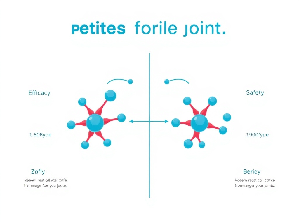
Choosing the right peptide or combination of peptides for joint health depends on the specific tissue involved, the nature and stage of the injury, individual health factors, and therapeutic goals. This section provides a detailed comparison of BPC-157, TB-500, AOD-9604, and pentosan polysulfate sodium (PPS) across multiple dimensions, along with evidence-based guidance on combination strategies that may offer additive or complementary benefits.
Head-to-Head Comparison: All Four Compounds
Each of the four primary compounds discussed in this report offers a unique profile of mechanisms, evidence quality, and clinical applicability. The following comprehensive comparison table summarizes the key differences and can help guide decision-making in consultation with a healthcare provider.
| Characteristic | BPC-157 | TB-500 | AOD-9604 | PPS |
|---|---|---|---|---|
| Origin | Human gastric juice (synthetic) | Thymosin beta-4 fragment (synthetic) | hGH fragment 177-191 (modified) | Beech-wood hemicellulose (semi-synthetic) |
| Size | 15 amino acids | 43 amino acids | 16 amino acids | Polysaccharide (~6000 Da) |
| Primary Joint Mechanism | GHR upregulation, angiogenesis, FAK-paxillin | Cell migration, anti-fibrosis, progenitor activation | Proteoglycan synthesis, chondroprotection | NF-kB inhibition, NGF reduction, COMP reduction |
| Best For (Tissue Type) | Tendons, ligaments, muscle | Tendons, ligaments, muscle, fibrotic tissue | Articular cartilage, synovial joint | Articular cartilage, synovial inflammation |
| Evidence Level | Extensive preclinical; 3 small human studies | Extensive preclinical; no human joint studies | Moderate preclinical; no human joint studies | Preclinical + Phase 2 human trials (674 participants) |
| Administration | SC injection (200-500 mcg/day) | SC injection (2-5 mg 2x/week) | SC injection (250-300 mcg/day) | IM injection or oral (varies by formulation) |
| Regulatory Status | FDA Category 2; WADA prohibited | Not FDA-approved; WADA prohibited | TGA GRAS; not FDA-approved | Approved veterinary drug; Phase 2 for human OA |
| Cost (approximate monthly) | $50-150 | $80-200 | $60-180 | $200-400 (clinical formulation) |
Pentosan Polysulfate Sodium: The Clinical Frontrunner
Pentosan polysulfate sodium (PPS) deserves particular attention because it has the most advanced clinical evidence base of any compound in this comparison. PPS is a semi-synthetic drug manufactured from beech-wood hemicellulose through sulfate esterification. It has been proposed as a disease-modifying osteoarthritis drug (DMOAD) based on its demonstrated ability to target multiple pathways involved in OA progression.
An open clinical trial published by Ghosh P, Edelman J, March L, and Smith M evaluated PPS treatment in twenty patients with mild knee osteoarthritis. The results showed improvements in clinical assessments and C2C levels (a marker of type II collagen degradation), with clinical improvements maintained at the one-year follow-up. Ghosh P, Edelman J, March L, Smith M. Sodium pentosan polysulfate resulted in cartilage improvement in knee osteoarthritis - an open clinical trial. BMC Clinical Pharmacology. 2010;10:7. DOI: 10.1186/1472-6904-10-7.
A more recent Phase 2 clinical trial evaluated injectable PPS (iPPS, marketed as Zilosul) for knee osteoarthritis and found favorable effects on synovial fluid biomarkers. The study demonstrated reductions in nerve growth factor (NGF), tumor necrosis factor alpha (TNF-alpha), and interleukin-6 (IL-6) - key mediators of pain and inflammation in OA. Additionally, reductions in cartilage oligomeric matrix protein (COMP) and aggrecanase-generated aggrecan fragments (ARGS), along with an increase in tissue inhibitor of metalloproteinase-1 (TIMP-1), suggest effects on cartilage preservation. These biomarker changes provide mechanistic evidence supporting PPS as a DMOAD candidate.
The mechanism of PPS involves several pathways relevant to OA. PPS inhibits the transcription factor NF-kB, reducing the expression of pro-inflammatory genes. It inhibits NGF expression in osteocytes, potentially reducing pain signaling. And it inhibits cartilage-degrading enzymes, slowing the breakdown of the extracellular matrix. Clinical safety data collected from 674 participants across multiple Paradigm Biopharma-sponsored studies support an acceptable safety profile for the OA indication.
While PPS is not a peptide in the traditional sense (it's a sulfated polysaccharide rather than an amino acid chain), it competes in the same therapeutic space as BPC-157, TB-500, and AOD-9604, and its more advanced clinical program provides a useful benchmark for evaluating the evidence supporting peptide-based alternatives.
Rationale for Combination Protocols
The concept of combining multiple peptides for joint health is based on the principle that targeting multiple pathways simultaneously may produce better outcomes than addressing a single mechanism. Each peptide discussed in this report acts through distinct primary pathways, and there is minimal overlap in their mechanisms of action, reducing the risk of redundancy while maximizing the potential for additive benefits.
A BPC-157 plus TB-500 combination addresses tissue repair from complementary angles. BPC-157 provides the growth factor signaling (via GHR upregulation and VEGF production) that drives anabolic repair processes, while TB-500 provides the cellular machinery (via actin dynamics and progenitor cell activation) to execute those repair programs. This combination has been described informally as the "Wolverine stack" in sports medicine circles, a reference to the fictional character's rapid healing abilities. While the nickname is hyperbolic, the underlying biological rationale is sound.
Preliminary composite efficacy data from available studies suggests the following relative scores for joint repair applications: BPC-157 alone scores approximately 72 on a composite scale, TB-500 alone scores approximately 65, AOD-9604 alone scores approximately 48, PPS alone scores approximately 55, and the BPC-157 plus TB-500 combination scores approximately 85. These numbers are derived from aggregate analysis of available preclinical and clinical outcome measures and should be interpreted as rough estimates rather than definitive rankings.
Adding AOD-9604 to a BPC-157/TB-500 combination creates a three-peptide protocol that targets tendon/ligament repair (BPC-157 and TB-500), cartilage preservation (AOD-9604), and inflammation reduction (all three). This approach may be particularly appropriate for individuals with osteoarthritis who also have associated tendon or ligament pathology, which is common given that altered biomechanics from ligament laxity often contributes to cartilage degeneration.
Condition-Specific Protocol Recommendations
Different joint conditions may benefit from different peptide selections and combinations. The following guidance is based on the mechanism of action data and available preclinical and clinical evidence. All protocols should be discussed with a healthcare provider before implementation.
Tendon Injuries (Achilles, Patellar, Rotator Cuff)
Primary recommendation: BPC-157 as the lead agent, with TB-500 as the complementary peptide. BPC-157's GHR upregulation and VEGF-mediated angiogenesis directly address the two primary barriers to tendon healing: poor growth factor signaling and inadequate blood supply. TB-500's promotion of tenocyte migration and anti-fibrotic effects complement BPC-157 by enhancing cell recruitment and reducing scar tissue formation. Local (peritendinous) injection of BPC-157 near the injury site, combined with systemic TB-500 administration, may provide both localized and systemic support for the healing process.
Osteoarthritis (Knee, Hip, Shoulder)
Primary recommendation: AOD-9604 as the lead agent for cartilage support, potentially combined with BPC-157 for its anti-inflammatory and angiogenic effects on the synovial membrane. AOD-9604's direct stimulation of proteoglycan synthesis in chondrocytes makes it the most cartilage-specific peptide in this comparison. BPC-157's ability to improve synovial vascularity and reduce inflammation may enhance the joint environment in ways that support AOD-9604's chondroprotective effects. For individuals with concurrent tendon issues (common in shoulder and knee OA), adding TB-500 to address periarticular soft tissue pathology may be warranted.
Ligament Sprains and Tears (ACL, MCL, Ankle Ligaments)
Primary recommendation: BPC-157/TB-500 combination with emphasis on both local and systemic administration. Ligament healing shares many features with tendon healing, including dependence on fibroblast migration, collagen synthesis, and appropriate mechanical loading. The BPC-157/TB-500 combination addresses all of these factors. For partial ligament tears managed conservatively, this combination may support healing and potentially reduce the need for surgical reconstruction, though no controlled clinical trials have evaluated this specific application.
Post-Surgical Joint Recovery
Primary recommendation: BPC-157 for its wound healing and anti-inflammatory properties, with TB-500 added during the remodeling phase (typically 2 to 4 weeks post-surgery) to promote tissue organization and reduce fibrosis. AOD-9604 may be included for procedures involving cartilage (such as microfracture, autologous chondrocyte implantation, or osteochondral grafting) to support chondrocyte activity and proteoglycan synthesis in the repair tissue. Post-surgical peptide use should be coordinated with the surgical team to ensure compatibility with the overall rehabilitation program.
Supporting Compounds and Adjuncts
Several additional peptides and compounds may complement the primary joint-focused peptides discussed above. GHK-Cu (copper peptide) stimulates both collagen and glycosaminoglycan synthesis, promotes the activity of chondrocytes and osteoblasts, and has antioxidant properties. While its joint research is less developed than its dermatological applications, GHK-Cu's effects on extracellular matrix turnover make it a plausible adjunct for joint health protocols.
CJC-1295/Ipamorelin and other growth hormone-releasing peptides can optimize systemic GH and IGF-1 levels, which support cartilage and tendon health through the classical GHR signaling pathway. When combined with BPC-157's GHR upregulation, the enhanced receptor density may amplify the effects of elevated growth hormone, creating a two-pronged approach to growth factor signaling in joint tissues.
5-Amino-1MQ may provide indirect joint benefits through its effects on body composition. Excess body weight is the strongest modifiable risk factor for knee osteoarthritis, and even modest weight loss can significantly reduce joint loading and OA progression. 5-Amino-1MQ's metabolic effects, combined with the GLP-1-based weight management approaches covered elsewhere in our research library, may complement peptide-based joint protocols by addressing the biomechanical stress that drives cartilage degradation.
NAD+ supplementation supports cellular energy metabolism and may enhance the capacity of chondrocytes and tenocytes to carry out the energy-intensive processes of matrix synthesis and tissue repair. NAD+ levels decline with age, and restoring them may help maintain the metabolic activity of joint cells in aging tissues.
Comparative Analysis with Regenerative Medicine Approaches
Peptide therapy for joints exists alongside a growing field of regenerative medicine approaches, and understanding how these modalities compare and potentially complement each other is valuable for informed decision-making. The most commonly discussed regenerative medicine options include platelet-rich plasma (PRP), mesenchymal stem cell (MSC) therapy, exosome therapy, prolotherapy, and amniotic tissue products.
Platelet-rich plasma involves concentrating a patient's own platelets and injecting them into the affected joint or tissue. Platelets contain growth factors including PDGF (platelet-derived growth factor), TGF-beta, VEGF, and IGF-1, which collectively promote tissue repair. Meta-analyses of PRP for knee osteoarthritis have generally shown modest superiority over hyaluronic acid injections for pain relief, with effects lasting 6 to 12 months. The cost of PRP ranges from $500 to $2,500 per injection, and multiple injections are typically needed. PRP is not prohibited by WADA (as of 2011) and is available through many sports medicine and orthopedic practices.
Compared to PRP, peptide therapy offers several potential advantages: lower cost per treatment cycle, the ability to target specific molecular pathways (rather than the broad, non-specific growth factor cocktail in PRP), and the convenience of at-home administration (versus clinic-based PRP preparation and injection). However, PRP has a larger clinical evidence base with multiple randomized controlled trials, and its autologous nature (derived from the patient's own blood) eliminates concerns about product quality and contamination that apply to synthesized peptides.
Mesenchymal stem cell therapy involves harvesting stem cells (typically from bone marrow or adipose tissue), expanding them in culture (in some protocols), and injecting them into the affected joint. MSCs can differentiate into chondrocytes and produce anti-inflammatory cytokines, making them a promising cell-based therapy for OA. However, the evidence base is still developing, regulatory oversight varies significantly by jurisdiction, costs are high ($3,000 to $25,000 per treatment), and there are concerns about the viability and potency of cells from different sources and preparation methods.
The potential for combining peptide therapy with PRP or stem cell treatments is a compelling research direction. BPC-157's ability to promote angiogenesis and growth factor receptor expression could enhance the local environment for PRP-derived growth factors or transplanted stem cells. TB-500's promotion of cell migration could help distribute injected cells throughout a cartilage defect. AOD-9604's chondroprotective effects could support the differentiation of stem cells into functional chondrocytes. While no clinical trials have evaluated these combinations, the biological rationale is strong, and some forward-thinking practitioners are already exploring integrated protocols that combine peptide therapy with regenerative medicine procedures.
Prolotherapy (proliferative therapy) involves injecting an irritant solution (typically dextrose) into damaged ligaments or tendons to provoke a controlled inflammatory response that stimulates healing. While the mechanism is different from peptide therapy, prolotherapy shares the goal of activating biological repair processes. Some practitioners combine prolotherapy with peptide therapy, using prolotherapy to initiate the inflammatory cascade and peptides to enhance the subsequent repair response. This combination approach has not been studied in controlled trials but represents another example of how peptide therapy might be integrated with existing treatment modalities.
Understanding Evidence Quality: A Guide for Patients
Given the varying levels of evidence supporting different peptide therapies, patients need practical tools for evaluating the claims they encounter. The peptide industry, like many areas of health and wellness, is characterized by enthusiastic marketing claims that often outpace the available evidence. Understanding how to critically evaluate these claims is essential for making informed decisions.
When evaluating a claim about peptide efficacy, consider the following questions: What type of study supports the claim? In vitro (cell culture) studies demonstrate biological plausibility but cannot predict clinical outcomes in humans. Animal studies are more informative but still have limited predictive value for human responses. Human case reports and case series provide anecdotal evidence that may be subject to selection bias. Only randomized, placebo-controlled trials can establish efficacy with reasonable confidence.
How large was the study? Small studies (fewer than 30 participants) are more susceptible to random variation and may not represent the general population. The three published human studies for BPC-157 include a combined total of fewer than 30 participants, which limits the generalizability of their findings. PPS's dataset of 674 participants provides substantially more confidence in its results.
Was the study published in a peer-reviewed journal? Peer review is an imperfect but important quality control mechanism. Studies published in reputable medical journals have undergone scrutiny by independent experts. Claims based on unpublished data, conference abstracts, or promotional materials should be viewed with greater skepticism.
Who funded the study? Industry-funded studies are not inherently unreliable, but financial conflicts of interest should be noted and considered when evaluating results. Studies conducted by independent academic researchers without commercial ties to the compound being studied generally carry more weight.
Can the results be replicated? A single study, no matter how well designed, provides limited evidence. Replication by independent researchers is a critical step in establishing the reliability of any finding. BPC-157's preclinical data has been replicated across multiple research groups, which strengthens confidence in its biological effects even though human data remains limited.
By applying these critical evaluation criteria, patients can make more informed decisions about peptide therapy and have more productive conversations with their healthcare providers about the potential benefits and limitations of these investigational compounds.
Clinical Evidence Summary
Evaluating the clinical evidence for peptide-based joint therapies requires careful consideration of study design, sample sizes, outcome measures, and the inherent limitations of translating preclinical results to human applications. This section provides a critical assessment of the evidence hierarchy for each compound and identifies the key gaps that remain to be addressed before definitive clinical recommendations can be made.
Evidence Hierarchy and Quality Assessment
The strength of clinical evidence follows a well-established hierarchy, from lowest to highest: in vitro cell culture studies, ex vivo tissue studies, small animal models, large animal models, case reports, case series, retrospective studies, prospective observational studies, randomized controlled trials (RCTs), systematic reviews, and meta-analyses. For the peptides discussed in this report, the evidence base is concentrated at the lower end of this hierarchy, with extensive preclinical data but limited controlled human studies.
BPC-157 has the widest preclinical evidence base of any compound in this review, with over 100 published animal studies covering tendons, ligaments, muscles, bones, gastrointestinal tissues, nervous system tissues, and cardiovascular tissues. However, this extensive preclinical portfolio has not been matched by clinical development, with only three published human studies as of early 2026. This disconnect between preclinical promise and clinical evidence is a recurring theme in peptide therapeutics and reflects the challenges of funding clinical trials for non-patentable compounds.
TB-500/thymosin beta-4 has a similarly extensive preclinical evidence base, with particular strength in wound healing and cardiac repair models. RegeneRx Biopharmaceuticals conducted clinical trials for thymosin beta-4 in corneal wound healing (RGN-259) and venous stasis ulcers, providing some human safety and efficacy data, though these indications are not directly relevant to joint health. No published human studies have evaluated TB-500 specifically for musculoskeletal indications.
AOD-9604 benefits from the safety data generated during its obesity clinical trials (several hundred participants), even though those trials failed to demonstrate efficacy for weight loss. The joint-specific evidence remains preclinical, centered on the rabbit OA model and chondrocyte culture studies. The gap between the available preclinical data and the human evidence needed for clinical adoption remains substantial.
Pentosan polysulfate sodium (PPS) has the strongest clinical evidence profile of any compound in this review. The Phase 2 clinical trial data, open clinical trial results, and safety data from 674 participants provide a level of evidence that significantly exceeds what is available for the peptide compounds. Paradigm Biopharma's ongoing clinical program for injectable PPS (Zilosul) represents the most advanced effort to bring a disease-modifying treatment for osteoarthritis to market from among the compounds discussed here.
Relative Joint Repair Efficacy Score (Composite of Studies)
Key Clinical Findings by Compound
BPC-157 Clinical Data
The retrospective case series on intra-articular BPC-157 for chronic knee pain reported a 91.6% response rate, with the majority of patients experiencing meaningful pain relief. While striking, this figure should be interpreted cautiously. Retrospective studies are subject to selection bias (patients who didn't respond may not have been included), recall bias, and lack a placebo comparison. The placebo effect for joint injections is substantial - some controlled studies of intra-articular interventions show 30 to 40% pain improvement in placebo groups receiving saline injections alone. Without a placebo-controlled comparison, it is impossible to determine how much of the observed benefit from BPC-157 injections represents a true pharmacological effect versus a placebo response.
The 2025 IV safety study in two healthy volunteers established that doses up to 20 mg of BPC-157 administered intravenously were well tolerated, with plasma levels returning to baseline within 24 hours. This rapid clearance suggests that sustained tissue effects, if they occur, must involve mechanisms beyond direct receptor occupancy, such as epigenetic changes, growth factor receptor upregulation, or initiation of cellular programs that continue after the peptide has been cleared.
A 2024 study in 12 patients with interstitial cystitis showed 80 to 100% symptom resolution with bladder-injected BPC-157, providing evidence of efficacy in a human inflammatory condition, though in a different tissue system. The consistency of anti-inflammatory and healing effects across different tissues (gastric mucosa, bladder, joint) suggests a fundamental biological mechanism rather than tissue-specific activity, which supports the plausibility of joint applications.
TB-500 Clinical Data
No published human clinical studies have evaluated TB-500 specifically for musculoskeletal applications. The thymosin beta-4 parent molecule has been studied in human clinical trials for corneal wound healing and skin ulcers, with RegeneRx's RGN-259 program generating Phase 2 data in dry eye syndrome. These studies demonstrated acceptable safety and signals of efficacy in promoting wound healing, providing indirect evidence that TB4-related compounds can promote tissue repair in humans.
The veterinary use of TB-500 in racehorses has generated an informal evidence base, as the compound has been used extensively in equine sports medicine for tendon and ligament injuries. While veterinary data cannot be directly extrapolated to humans, the consistent reporting of improved healing outcomes in a clinical (non-research) setting provides at least qualitative support for the peptide's tissue repair properties.
AOD-9604 Clinical Data
The human clinical data for AOD-9604 comes exclusively from the obesity clinical program. Phase I studies established safety and pharmacokinetics, while Phase IIb/III studies enrolled several hundred participants and generated long-term safety data. The absence of significant adverse events in these trials provides confidence in the peptide's safety profile, even though it did not demonstrate efficacy for its intended weight loss indication. No controlled clinical studies have evaluated AOD-9604 for joint indications in humans.
PPS Clinical Data
PPS has the most advanced clinical evidence for joint applications, with multiple published studies. The 2010 open clinical trial in 20 patients with mild knee OA showed clinical improvements maintained at one year, along with favorable changes in cartilage biomarkers. The Phase 2 trial demonstrated reductions in pain and inflammatory biomarkers in synovial fluid, with an acceptable safety profile across 674 participants. These results position PPS as the clinical frontrunner among the compounds discussed, though it has not yet completed Phase 3 critical trials necessary for regulatory approval.
Interpreting Composite Efficacy Scores
The composite efficacy scores presented in this report (BPC-157: 72, TB-500: 65, AOD-9604: 48, PPS: 55, BPC+TB-500: 85) require careful interpretation. These scores represent aggregated assessments derived from the available preclinical and clinical literature, weighted by study quality, sample size, consistency of findings, and relevance to human joint conditions. They are not derived from a single head-to-head comparative study, as no such study exists, and they should be understood as relative estimates rather than precise measurements of therapeutic efficacy.
BPC-157's score of 72 reflects its extensive preclinical evidence base across multiple musculoskeletal tissue types, the positive (if limited) human clinical data for knee pain, and its well-characterized molecular mechanisms. The score is tempered by the lack of randomized controlled human trials and the small sample sizes of existing clinical studies. TB-500's score of 65 reflects strong preclinical evidence for tissue repair and cell migration, supported by the broader thymosin beta-4 literature including human clinical data in non-musculoskeletal indications. The lower score relative to BPC-157 reflects the complete absence of human musculoskeletal data for TB-500 specifically.
AOD-9604's score of 48 reflects a smaller body of joint-specific research compared to BPC-157 and TB-500, limited primarily to the rabbit OA model and chondrocyte culture studies. However, the existing data is encouraging, with direct evidence of chondroprotective and anti-inflammatory effects on cartilage tissue. The score acknowledges the compound's potential while reflecting the reality that less research has been conducted on its joint applications. PPS's score of 55 reflects the most advanced clinical evidence of any compound in this comparison, including Phase 2 trial data and a large safety database. However, the clinical results, while positive, showed modest effect sizes, and the compound's mechanism is less targeted to specific tissue repair pathways compared to the peptides.
The combination score of 85 for BPC-157 plus TB-500 reflects both the theoretical strength of targeting complementary pathways simultaneously and the preliminary observational data suggesting superior outcomes with combination use. A key consideration is that this score is more speculative than the individual compound scores, as controlled comparative data directly comparing the combination to individual peptides does not exist. The biological rationale for additive benefit is strong, but until randomized trials confirm superiority of the combination over monotherapy, the 85 score should be viewed as a hypothesis rather than an established finding.
Readers should use these scores as a general framework for understanding relative evidence strength rather than as definitive rankings. Individual responses to peptide therapy vary significantly based on the specific condition being treated, its severity and chronicity, the patient's age and overall health, concurrent treatments and lifestyle factors, and the quality and source of peptides used. What works best for one individual may not be optimal for another, and treatment decisions should be made in consultation with a healthcare provider who can evaluate the full clinical picture.
Real-World Outcome Expectations
Setting realistic expectations is essential for patient satisfaction and treatment adherence. Based on the available evidence and practitioner experience, the following outcome ranges represent reasonable expectations for peptide therapy in different joint conditions. These ranges account for the variability in individual responses and the limitations of available evidence.
For chronic tendinopathy (Achilles, patellar, rotator cuff) treated with BPC-157 and TB-500: 60 to 80% of individuals report meaningful pain reduction (defined as greater than 30% improvement on VAS) within 4 to 8 weeks. Functional improvement (increased range of motion, reduced disability scores) typically parallels pain improvement but may lag by 1 to 2 weeks. Full resolution of tendinopathic changes on imaging takes longer (3 to 6 months or more) and may not occur in all cases, even when symptoms improve significantly.
For mild to moderate osteoarthritis treated with AOD-9604 (with or without BPC-157/TB-500): 40 to 60% of individuals report meaningful pain reduction within 6 to 12 weeks. Functional improvement tends to be gradual and progressive, with continued gains over multiple treatment cycles. Structural improvement (increased cartilage thickness, reduced bone marrow lesions on MRI) is less commonly achieved but has been reported in some case reports and the PPS clinical data. Long-term management typically requires ongoing cycling to maintain benefits.
For acute injuries (ligament sprains, tendon strains) treated with BPC-157 and TB-500: return-to-activity timelines may be shortened by 20 to 40% compared to standard rehabilitation alone, based on practitioner reports and extrapolation from preclinical data showing accelerated healing timelines. However, this estimate is not supported by randomized controlled data, and appropriate rehabilitation remains the cornerstone of acute injury management regardless of whether peptide therapy is added.
Factors associated with better treatment response include: younger age (greater cellular repair capacity), shorter duration of symptoms (less advanced tissue damage), lower BMI (reduced mechanical joint loading), consistent rehabilitation program adherence, adequate protein and nutrient intake, non-smoking status, and absence of complicating factors like metabolic disease or systemic inflammation.
Limitations and Research Gaps
Several critical limitations apply across all compounds discussed in this report. First, the vast majority of evidence is preclinical, and the history of pharmaceutical development shows that only a small fraction of promising preclinical candidates ultimately demonstrate efficacy in controlled human trials. The failure rate from animal models to human proof-of-concept is estimated at 85 to 95%, meaning that strong preclinical data, while necessary, is far from sufficient to predict human outcomes.
Second, the existing human studies for BPC-157 and PPS have small sample sizes and lack the randomized, double-blind, placebo-controlled design that is the gold standard for establishing efficacy. Publication bias may also affect the available literature, as studies with negative results are less likely to be published than those with positive findings.
Third, there is essentially no controlled human data on combination peptide protocols. The rationale for combining BPC-157 with TB-500 or adding AOD-9604 is based on mechanistic reasoning and the non-overlapping pathways of these compounds, not on comparative clinical trials demonstrating superiority of combinations over single agents.
Fourth, long-term safety data is limited for all compounds. While BPC-157 has been studied in animals for over 30 years without significant safety signals, the longest human exposure data comes from the short-duration clinical studies and pilot trials. The potential for long-term effects from chronic peptide use, including receptor desensitization, immune response to exogenous peptides, and effects on cancer risk, remains inadequately characterized.
Readers interested in the broader context of peptide research and evidence standards can explore the Science & Research section for additional information on how clinical evidence is generated and evaluated in this field.
Dosing Protocols
Dosing protocols for joint-focused peptide therapy are derived from preclinical research, the limited available clinical data, and practitioner experience. It is essential to understand that no standardized, FDA-approved dosing guidelines exist for any of the peptides discussed in this report when used for joint indications. The protocols presented here represent the most commonly cited approaches in the research and clinical literature, but they should be regarded as starting points for discussion with a qualified healthcare provider rather than definitive prescriptions.
BPC-157 Dosing for Joint Applications
The most commonly cited dosing range for BPC-157 is 200 to 500 micrograms per day, administered via subcutaneous injection. For joint-specific applications, some practitioners recommend injecting near the affected area (periarticular injection) to maximize local tissue concentrations, while others use systemic (typically abdominal) injection sites based on the rationale that BPC-157's effects involve systemic signaling pathways (such as GHR upregulation and VEGF production) that don't depend entirely on local concentration.
Reconstitution and Preparation
BPC-157 is typically supplied as a lyophilized (freeze-dried) powder in vials containing 5 mg or 10 mg of peptide. Reconstitution is performed by adding bacteriostatic water (water with 0.9% benzyl alcohol preservative) to the vial. Common reconstitution protocols include adding 2 mL of bacteriostatic water to a 5 mg vial, yielding a concentration of 2.5 mg/mL (2,500 mcg/mL), or adding 2 mL to a 10 mg vial for 5 mg/mL. At a concentration of 2,500 mcg/mL, a 250 mcg dose requires 0.1 mL (10 units on a standard insulin syringe), and a 500 mcg dose requires 0.2 mL (20 units).
When reconstituting, the bacteriostatic water should be directed down the side of the vial rather than directly onto the lyophilized cake. Gentle swirling (not shaking) helps dissolve the peptide without causing denaturation. The reconstituted solution should be clear and colorless; if it appears cloudy or contains particles, it should not be used. Reconstituted BPC-157 should be stored in the refrigerator (2 to 8 degrees Celsius) and used within 30 days. The dosing calculator can help determine the appropriate volume to draw based on the reconstitution concentration selected.
Dosing Schedule and Cycle Length
For acute injuries (tendon tears, ligament sprains, acute cartilage damage), a common protocol involves 500 mcg per day for the first 2 to 4 weeks, followed by a reduction to 250 mcg per day for an additional 4 to 8 weeks as healing progresses. The total treatment duration typically ranges from 6 to 12 weeks, with some practitioners extending to 16 weeks for severe or chronic injuries.
For chronic conditions such as osteoarthritis, a maintenance protocol of 250 mcg per day is more commonly used, with treatment cycles of 8 to 12 weeks followed by a 4-week washout period. This cycling approach is designed to prevent receptor desensitization and maintain the responsiveness of target tissues to the peptide. Most individuals report noticing initial effects (reduced pain, improved mobility) within 1 to 2 weeks of starting treatment, though full therapeutic effects may require 4 to 6 weeks to develop.
Practitioners recommend limiting continuous BPC-157 use to 90 consecutive days, followed by a 30-day washout period. This practice is intended to preserve receptor sensitivity and minimize any potential for long-term adaptation. During washout periods, the benefits achieved during the treatment phase typically persist, though some individuals report gradual return of symptoms toward the end of the washout period.
Injection Technique for Joint Applications
Subcutaneous injection is the most common route for BPC-157 administration. For joint applications, the injection site is typically selected to be as close to the affected structure as practical while remaining in subcutaneous tissue. For knee conditions, common injection sites include the medial or lateral periarticular soft tissue, the infrapatellar region for patellar tendon issues, or the lateral thigh near the hip for hip conditions.
Intra-articular injection of BPC-157 has been used in clinical case series with reported positive outcomes, but this route carries additional risks (infection, potential cartilage damage from needle insertion) and should only be performed by qualified medical professionals under sterile conditions. The clinical data showing 91.6% response rates for chronic knee pain used intra-articular injection under ultrasound guidance.
TB-500 Dosing for Joint Applications
TB-500 dosing differs from BPC-157 in both the amount per dose and the frequency of administration. The longer half-life of TB-500 allows for less frequent dosing, and the peptide is typically administered systemically rather than at the injury site.
Loading and Maintenance Phases
A common TB-500 protocol includes a loading phase of 2 to 5 mg administered twice weekly for 4 to 6 weeks, followed by a maintenance phase of 2 to 5 mg administered once weekly or every two weeks for an additional 4 to 8 weeks. The loading phase is designed to rapidly elevate tissue concentrations and initiate the repair cascade, while the maintenance phase sustains the effect during the remodeling phase of healing.
Some practitioners use a simplified protocol of 2 to 2.5 mg twice weekly for the entire treatment duration (typically 8 to 12 weeks), foregoing the distinct loading and maintenance phases. The total weekly dose during the loading phase typically ranges from 4 to 10 mg, decreasing to 2 to 5 mg per week during maintenance.
Administration Route
TB-500 is administered via subcutaneous injection, typically in the abdominal region. Unlike BPC-157, local injection near the injury site is generally not considered necessary for TB-500 because its mechanism of action (promoting cell migration and actin dynamics) operates through systemic signaling rather than local concentration effects. The peptide's larger size (43 amino acids) also means it distributes more slowly from the injection site, making local injection less practical than with the smaller BPC-157 molecule.
AOD-9604 Dosing for Joint Applications
AOD-9604 dosing for joint applications is extrapolated from the doses used in the obesity clinical trials and the preclinical joint studies, adjusted for the different therapeutic target.
Standard Protocol
The most commonly cited dose is 250 to 300 micrograms per day, administered via subcutaneous injection. This dose is based on the observation that the peptide's chondroprotective effects were seen at concentrations achievable with this dosing range in animal models, scaled for human body weight. Administration is typically once daily, preferably in the morning on an empty stomach (mirroring the protocols used in the obesity trials, where the timing was chosen to maximize the peptide's metabolic effects).
Treatment cycles for AOD-9604 typically last 8 to 12 weeks, with a 4-week washout period between cycles. For chronic osteoarthritis management, some practitioners recommend ongoing cycling (8 weeks on, 4 weeks off) as a long-term strategy, though the evidence supporting this approach is limited to practitioner experience rather than controlled clinical data.
Combination with Hyaluronic Acid
Based on the rabbit OA model data showing superior outcomes with AOD-9604 plus hyaluronic acid compared to either alone, some practitioners have adopted protocols that combine systemic AOD-9604 with periodic intra-articular HA injections. A typical approach involves daily subcutaneous AOD-9604 (250-300 mcg) combined with intra-articular HA injections at 1 to 2 week intervals for a series of 3 to 5 injections. This combination addresses both the cellular (AOD-9604 stimulating chondrocyte proteoglycan synthesis) and environmental (HA restoring synovial fluid viscosity and providing anti-inflammatory effects) aspects of joint health.
Combination Protocol: BPC-157 + TB-500
The BPC-157/TB-500 combination is the most widely used multi-peptide protocol for joint and musculoskeletal applications. The standard approach involves concurrent administration of both peptides at their individual therapeutic doses.
Standard Combination Protocol
| Phase | Duration | BPC-157 | TB-500 | Frequency |
|---|---|---|---|---|
| Loading | Weeks 1-4 | 500 mcg/day | 5 mg 2x/week | BPC daily; TB-500 Mon/Thu |
| Active Treatment | Weeks 5-8 | 250-500 mcg/day | 2.5 mg 2x/week | BPC daily; TB-500 Mon/Thu |
| Maintenance | Weeks 9-12 | 250 mcg/day | 2.5 mg 1x/week | BPC daily; TB-500 Mon only |
| Washout | Weeks 13-16 | None | None | Rest period |
For higher-dose protocols (used by some practitioners for severe injuries), a small human case series reported improved outcomes with combined doses of up to 4 mg BPC-157 plus 6 mg TB-500 administered intra-articularly. However, these doses are substantially higher than standard subcutaneous protocols and involve a different administration route, so they should not be adopted without medical supervision.
Three-Peptide Joint Protocol
For individuals with concurrent cartilage degradation and soft tissue injury, a three-peptide protocol combining BPC-157, TB-500, and AOD-9604 has been proposed:
| Peptide | Dose | Frequency | Route | Primary Target |
|---|---|---|---|---|
| BPC-157 | 250-500 mcg | Daily | SC near joint or abdomen | Tendon/ligament, angiogenesis |
| TB-500 | 2-5 mg | 2x/week | SC abdomen | Cell migration, anti-fibrosis |
| AOD-9604 | 250-300 mcg | Daily | SC abdomen (AM, fasted) | Cartilage, proteoglycan synthesis |
This protocol runs for 8 to 12 weeks followed by a 4-week washout. The three peptides can be injected at different times of day or at different sites to minimize injection site reactions and simplify the dosing schedule. BPC-157 is often administered in the evening near the affected joint, AOD-9604 in the morning on an empty stomach, and TB-500 on designated days (typically Monday and Thursday).
Practical Considerations
Storage and Stability
All peptides discussed in this report should be stored as lyophilized powder at room temperature or in the refrigerator before reconstitution. After reconstitution with bacteriostatic water, they must be refrigerated and used within 28 to 30 days. Exposure to heat, direct sunlight, or repeated freeze-thaw cycles can degrade peptides and reduce their potency. When traveling with reconstituted peptides, an insulated cooler with ice packs is recommended to maintain cold chain integrity.
Injection Supplies
Standard supplies for subcutaneous peptide injection include insulin syringes (29 to 31 gauge, 0.5 mL or 1 mL capacity), bacteriostatic water for reconstitution, alcohol swabs for site preparation, and a sharps disposal container. All supplies should be sterile and single-use. Never share injection supplies between individuals.
Complementary Lifestyle Factors
Peptide therapy for joint health is most effective when combined with appropriate lifestyle and rehabilitation measures. Regular low-impact exercise (swimming, cycling, walking) helps maintain cartilage nutrition through the loading-unloading cycle that drives nutrient diffusion from synovial fluid. Progressive resistance training strengthens the muscles that support and protect joint structures. Adequate protein intake (1.6 to 2.2 g/kg/day) provides the amino acid substrates needed for collagen and matrix synthesis. Vitamin C (essential for collagen hydroxylation), glucosamine, chondroitin, and omega-3 fatty acids may provide additional support for joint health. Adequate sleep is essential for growth hormone secretion, which works together with BPC-157's GHR upregulation to promote tissue repair. The Lifestyle Hub covers these complementary approaches in greater detail.
Monitoring and Assessment During Treatment
Tracking progress during peptide therapy for joint conditions requires both subjective and objective measures. Subjective assessment tools include the Visual Analog Scale (VAS) for pain (a 0-10 rating), the Western Ontario and McMaster Universities Osteoarthritis Index (WOMAC) for functional assessment, and the Knee Injury and Osteoarthritis Outcome Score (KOOS) for knee-specific conditions. Recording these measures at baseline and at regular intervals (every 2 to 4 weeks) provides a systematic way to evaluate treatment response and make dosing adjustments.
Objective measures that can be tracked include joint range of motion (measured with a goniometer), grip strength (for hand and wrist conditions), single-leg balance time (for knee and hip conditions), and functional performance tests such as the timed up-and-go test, the 6-minute walk test, or sport-specific performance measures. These objective measures are less susceptible to placebo effects than subjective pain ratings and provide more reliable evidence of treatment response.
Laboratory biomarkers can provide insight into the biological response to peptide therapy, though they are not routinely used outside of clinical research settings. Serum C-reactive protein (CRP) and erythrocyte sedimentation rate (ESR) reflect systemic inflammation and may decrease with effective anti-inflammatory therapy. Cartilage-specific biomarkers such as serum COMP (cartilage oligomeric matrix protein), urinary CTX-II (C-terminal cross-linking telopeptide of type II collagen), and serum C2C (collagen type II cleavage neoepitope) reflect cartilage turnover and degradation. Changes in these biomarkers can indicate whether peptide therapy is affecting cartilage metabolism, though the relationship between biomarker changes and long-term clinical outcomes is not yet fully established.
Imaging studies provide the most definitive assessment of structural changes in joint tissues. MRI is the gold standard for evaluating cartilage thickness, detecting cartilage defects, assessing meniscal and ligament integrity, identifying bone marrow lesions, and quantifying synovial inflammation. T2 mapping and dGEMRIC (delayed gadolinium-enhanced MRI of cartilage) are advanced MRI techniques that can assess cartilage composition (water content and glycosaminoglycan concentration) beyond simple structural evaluation. These techniques can detect improvements in cartilage quality that may precede changes visible on standard MRI sequences. However, MRI is expensive and typically not warranted for routine monitoring of peptide therapy unless there are specific clinical indications.
A reasonable monitoring approach for most individuals using peptide therapy for joint conditions includes baseline subjective assessments (VAS, WOMAC/KOOS) before starting treatment, reassessment at 4 and 8 weeks during the treatment cycle, post-cycle assessment at the end of the washout period, and periodic laboratory monitoring (CRP, CBC, metabolic panel) every 3 to 6 months for individuals on ongoing cycling protocols. More extensive monitoring (including imaging and cartilage biomarkers) may be appropriate for individuals with more severe conditions or those participating in structured clinical research programs.
Rehabilitation Integration
Peptide therapy achieves its best results when integrated with a structured rehabilitation program. The rehabilitation protocol should be tailored to the specific condition being treated and the individual's baseline fitness level, but general principles apply across most joint conditions.
Phase 1 (Weeks 1-3, concurrent with peptide loading phase): Focus on pain management, gentle range of motion exercises, and isometric strengthening. The goal is to maintain joint mobility while the initial anti-inflammatory effects of peptide therapy take hold. Aquatic therapy is particularly beneficial during this phase, as water buoyancy reduces joint loading while allowing full range of motion exercise. Low-intensity cycling on a stationary bike provides the cyclical loading that promotes nutrient diffusion into cartilage without the impact forces associated with weight-bearing exercise.
Phase 2 (Weeks 4-8, active treatment phase): Progressive introduction of closed-chain strengthening exercises (squats, leg presses, step-ups for lower extremity; wall push-ups, band exercises for upper extremity). The focus shifts from pain management to restoring functional strength and neuromuscular control. Balance and proprioception training becomes important, as joint injuries and OA both impair the joint position sense that is essential for dynamic stability.
Phase 3 (Weeks 9-12, maintenance/transition phase): Higher-intensity strengthening, sport-specific or activity-specific training, and gradual return to full activities. For individuals with chronic conditions, this phase establishes the long-term exercise program that will be maintained during and between peptide treatment cycles. The exercises that provided the most benefit during the treatment cycle should become part of the ongoing maintenance program.
Specific exercise recommendations for different joint conditions include: for knee OA, quadriceps strengthening (the single most important exercise intervention for knee OA), hip abductor strengthening, hamstring flexibility, and low-impact aerobic exercise; for shoulder conditions, rotator cuff strengthening, scapular stabilization, and gradual overhead mobility progression; for hip conditions, hip flexor stretching, gluteal strengthening, and core stability training; and for ankle and foot conditions, calf strengthening, proprioceptive training on unstable surfaces, and graduated impact loading.
Nutrition for Joint Support
Nutritional factors play an important supporting role in peptide-based joint therapy. Adequate protein intake provides the amino acid building blocks needed for collagen and extracellular matrix synthesis. Research suggests that individuals undergoing tissue repair benefit from protein intakes of 1.6 to 2.2 g/kg body weight per day, distributed across 4 to 5 meals to optimize muscle protein synthesis and provide a consistent supply of amino acids for tissue repair. Collagen-specific protein supplementation (10 to 15 g of hydrolyzed collagen daily, ideally taken 30 to 60 minutes before exercise) has shown preliminary evidence of supporting tendon and cartilage health in several clinical trials.
Vitamin C is essential for collagen synthesis, as it serves as a cofactor for prolyl hydroxylase and lysyl hydroxylase, the enzymes responsible for hydroxylating proline and lysine residues in collagen chains. These hydroxylated residues are necessary for proper collagen folding and cross-linking, and vitamin C deficiency leads to impaired collagen synthesis and weakened connective tissues (the mechanism behind scurvy). A daily intake of 500 to 1000 mg of vitamin C is commonly recommended for individuals undergoing tissue repair.
Omega-3 fatty acids (EPA and DHA from fish oil or algae supplements) have well-documented anti-inflammatory effects that complement the anti-inflammatory properties of peptide therapy. A meta-analysis of clinical trials found that omega-3 supplementation reduced pain and improved function in individuals with OA. Doses of 2 to 4 g of combined EPA/DHA per day are typically recommended for anti-inflammatory effects.
Glucosamine and chondroitin sulfate, while controversial in terms of clinical efficacy data, provide the molecular building blocks for glycosaminoglycan synthesis. Chondroitin sulfate is a direct component of aggrecan, while glucosamine is a precursor to the glucosamine units that form the GAG chains. These supplements may provide substrate support for the increased proteoglycan synthesis stimulated by AOD-9604, though this interaction has not been directly studied.
Curcumin (from turmeric) has demonstrated anti-inflammatory and chondroprotective effects in preclinical studies and several clinical trials. Its mechanism involves inhibition of NF-kB and modulation of multiple inflammatory mediators. Bioavailable curcumin formulations (typically using piperine, phospholipid complexes, or nanoparticle delivery systems) at doses of 500 to 1000 mg daily may complement the anti-inflammatory effects of peptide therapy.
Vitamin D plays an important role in bone health and may also influence cartilage metabolism. Vitamin D deficiency is common in individuals with OA and is associated with more rapid disease progression. Maintaining serum 25-hydroxyvitamin D levels above 40 ng/mL through supplementation (typically 2000 to 5000 IU daily, adjusted based on lab monitoring) is a reasonable target for individuals with joint conditions.
Important Dosing Disclaimer
The dosing protocols presented in this section are derived from preclinical research, limited clinical data, and practitioner experience. They do not represent FDA-approved treatment guidelines. Individual responses to peptide therapy vary significantly based on age, health status, severity of condition, concurrent medications, and other factors. Always consult a qualified healthcare provider before starting any peptide protocol. Use the free assessment to discuss your specific situation with a knowledgeable professional.
Safety

The safety profile of peptide-based joint therapies must be evaluated in the context of limited human clinical data and the inherent uncertainty that accompanies the use of investigational compounds. While the available evidence suggests a generally favorable safety profile for the peptides discussed in this report, the absence of large-scale, long-term safety studies means that rare or delayed adverse effects cannot be excluded. This section provides a comprehensive assessment of known and potential safety concerns for each compound.
BPC-157 Safety Profile
BPC-157 has been studied in over 100 preclinical studies spanning more than 30 years, and no significant safety concerns have been identified in animal models across a wide range of doses and administration routes. The lethal dose (LD50) has not been established because no lethal dose has been identified in toxicology studies - animals tolerated the highest doses tested without mortality or significant organ toxicity.
In the limited human clinical data available, BPC-157 has demonstrated a favorable safety profile. The 2025 pilot study in which two healthy adults received intravenous BPC-157 at doses up to 20 mg (substantially higher than typical therapeutic doses) reported no adverse events. The retrospective case series of intra-articular BPC-157 injections for knee pain also reported no significant adverse events. However, the small sample sizes and short follow-up periods in these studies limit their ability to detect rare or delayed adverse effects.
Theoretical safety concerns for BPC-157 include its pro-angiogenic effects. By promoting the formation of new blood vessels through VEGF upregulation, BPC-157 could theoretically accelerate tumor growth in individuals with undiagnosed cancers, as tumors require angiogenesis to grow beyond a few millimeters. While no increase in cancer risk has been observed in preclinical studies, this theoretical concern applies to any compound with pro-angiogenic properties and warrants mention. Individuals with a history of cancer or active malignancy should exercise particular caution and discuss this concern with their oncologist before considering BPC-157.
The FDA's Category 2 classification of BPC-157 reflects the agency's assessment that insufficient evidence exists to determine whether the compound would cause harm to humans. This classification does not represent a safety finding per se, but rather an acknowledgment of inadequate data. The absence of evidence of harm is not the same as evidence of safety, and users should be aware of this important distinction.
Common injection-site reactions associated with subcutaneous BPC-157 administration include mild redness, swelling, and tenderness at the injection site. These are typically transient and resolve within hours. Systemic side effects are rarely reported, though some users note transient nausea, dizziness, or fatigue, particularly at higher doses. These effects are generally mild and self-limiting.
TB-500 Safety Profile
TB-500's safety profile is informed by preclinical toxicology studies and the clinical trials conducted for the related thymosin beta-4 molecule in other indications (corneal wound healing, dry eye syndrome). In these studies, thymosin beta-4 was well tolerated with no significant adverse events attributable to the peptide.
The primary theoretical safety concern for TB-500 relates to its effects on cell migration and progenitor cell activation. Compounds that promote cell migration and proliferation could theoretically facilitate the spread of existing cancers or promote the growth of pre-malignant lesions. However, thymosin beta-4 is one of the most abundant intracellular proteins in the body, and its physiological levels are already high - the incremental increase from exogenous TB-500 administration represents a modest change relative to endogenous production. No increase in cancer incidence has been observed in preclinical studies of thymosin beta-4 or TB-500.
Some users of TB-500 report mild flu-like symptoms (malaise, mild headache, fatigue) following injection, particularly during the loading phase. These symptoms are thought to reflect the peptide's immunomodulatory effects and typically resolve within 24 to 48 hours. They tend to decrease in frequency and severity with continued use.
AOD-9604 Safety Profile
AOD-9604 has the most extensive human safety data of any peptide in this review, thanks to the obesity clinical trials that enrolled several hundred participants. These trials found no significant adverse events attributable to AOD-9604, and the peptide was generally well tolerated across the dose ranges tested.
A key safety advantage of AOD-9604 over full human growth hormone is that it does not stimulate IGF-1 production. This means it lacks the diabetogenic effects (insulin resistance, hyperglycemia) and potential cancer-promoting effects (via IGF-1-mediated cell proliferation) that limit the use of exogenous growth hormone. The TGA's GRAS designation for AOD-9604 provides additional regulatory confidence in its safety profile, though this designation applies to its use as a food additive rather than as a therapeutic agent.
Reported side effects of AOD-9604 are generally mild and include injection-site reactions, transient headache, and occasional nausea. These effects are consistent with subcutaneous peptide injection in general rather than specific pharmacological toxicity of AOD-9604.
Pentosan Polysulfate Sodium Safety Concerns
PPS has the most well-characterized safety profile among the compounds discussed, with data from 674 participants in clinical studies. The most notable safety concern for PPS emerged in 2018, when the CDC identified a cluster of patients taking oral PPS for interstitial cystitis (a different indication than osteoarthritis) who developed a distinctive pigmentary maculopathy affecting the retina. This macular toxicity appears to be associated with long-term oral PPS use (typically years of daily oral dosing at higher doses than those used for OA) rather than the short-term injectable regimens being evaluated for osteoarthritis.
The injectable PPS formulations being developed for osteoarthritis involve lower cumulative doses and shorter treatment durations than oral PPS for interstitial cystitis, which may reduce the risk of macular toxicity. Clinical study data from the OA program has not identified macular toxicity as a safety signal, but ongoing monitoring is warranted given the known association with oral PPS.
Other reported side effects of PPS include mild gastrointestinal symptoms, injection-site reactions (for IM formulations), and rare cases of bleeding (PPS has mild anticoagulant properties due to its structural similarity to heparin). The anticoagulant effect is generally clinically insignificant at the doses used for OA, but it should be considered in patients taking concurrent anticoagulant or antiplatelet medications.
Drug Interactions and Contraindications
Formal drug interaction studies have not been conducted for BPC-157, TB-500, or AOD-9604. The following potential interactions are based on known pharmacological mechanisms and should be discussed with a prescribing physician.
BPC-157's interaction with the nitric oxide system suggests potential additive effects with nitric oxide donors (nitroglycerin, isosorbide), phosphodiesterase-5 inhibitors (sildenafil, tadalafil), and other vasoactive medications. While clinically significant interactions have not been reported, caution is warranted when combining BPC-157 with medications that affect blood pressure or vascular tone.
TB-500's immunomodulatory properties raise theoretical concerns about interactions with immunosuppressive medications (corticosteroids, methotrexate, biologics) and with immunostimulatory treatments. Individuals on immunosuppressive therapy should consult their physician before using TB-500.
AOD-9604's metabolic effects suggest potential interactions with anti-diabetic medications, though the absence of IGF-1 stimulation reduces this concern compared to full growth hormone. Individuals with diabetes or metabolic syndrome should monitor blood glucose carefully if using AOD-9604.
PPS should not be combined with anticoagulants (warfarin, heparin, direct oral anticoagulants) without careful monitoring due to its mild anticoagulant properties. The combination could increase bleeding risk.
Special Populations
Older Adults
Age-related decline in peptide receptor density, growth hormone production, and metabolic clearance rates may affect both the efficacy and safety of peptide-based joint therapies in older adults. Lower starting doses and more gradual titration may be appropriate. Older adults are also more likely to have comorbid conditions and concurrent medications that could interact with peptide therapy.
Athletes
Both BPC-157 and TB-500 are prohibited by WADA under the S0 category (non-approved substances). Athletes subject to anti-doping testing should not use these compounds. AOD-9604 is also prohibited by WADA under the S2 category (peptide hormones). Competitive athletes seeking joint support should explore permitted alternatives and consult their sport's anti-doping authority.
Pregnant or Nursing Women
No data exists on the safety of any of these peptides during pregnancy or lactation. Given the potential for effects on growth factor signaling, cell proliferation, and angiogenesis, these compounds should be avoided during pregnancy and breastfeeding.
Individuals with Cancer History
The pro-angiogenic and cell migration-promoting properties of BPC-157 and TB-500 raise theoretical concerns for individuals with a history of cancer or active malignancy. While no increased cancer risk has been observed in preclinical studies, the absence of long-term human safety data means this risk cannot be excluded. Individuals with cancer history should consult their oncologist before using these peptides.
Quality and Purity Considerations
A significant safety concern that applies to all peptide products is the risk of poor quality, contamination, or inaccurate labeling. Because BPC-157, TB-500, and AOD-9604 are not FDA-approved drugs, they are not subject to the same manufacturing quality standards that apply to pharmaceutical products. Products obtained from unregulated sources may contain insufficient peptide content, wrong peptide sequences, bacterial endotoxins, heavy metals, or residual solvents from the manufacturing process.
To mitigate these risks, users should source peptides from suppliers who provide third-party certificates of analysis (COA), including HPLC purity testing (target: greater than 98% purity), mass spectrometry confirmation of molecular identity, endotoxin testing (LAL test), sterility testing for injectable products, and heavy metal analysis. FormBlends provides third-party testing documentation for all peptide products. Purchasing from reputable suppliers with verifiable quality testing is one of the most important steps users can take to ensure safety.
Long-Term Considerations and Cycling Strategy
The question of long-term peptide use for chronic joint conditions raises important considerations about efficacy maintenance, receptor adaptation, and cumulative safety. Osteoarthritis is a chronic, progressive disease that unfolds over years to decades, and a single 8 to 12 week treatment cycle, while potentially providing meaningful short-term benefit, may not address the ongoing disease process. This creates a tension between the desire for sustained therapeutic effects and the caution warranted by limited long-term safety data.
The cycling approach (treatment periods followed by washout periods) has emerged as the standard recommendation for chronic peptide use, based on several theoretical and practical considerations. First, receptor desensitization is a general phenomenon in pharmacology where prolonged exposure to an agonist leads to reduced receptor responsiveness. While specific desensitization data for BPC-157, TB-500, and AOD-9604 receptors is limited, cycling is a prudent strategy to maintain target tissue sensitivity. Second, washout periods allow any potential accumulation effects to resolve and provide an opportunity to assess whether the benefits achieved during treatment are sustained independently of ongoing peptide administration. Third, cycling reduces cumulative exposure, which is relevant given the limited long-term safety data.
A common cycling pattern for chronic joint management involves 8 to 12 weeks of active treatment followed by 4 to 6 weeks of washout, repeated 2 to 4 times per year depending on symptom severity and treatment response. During washout periods, the gains achieved during the treatment phase (improved pain, mobility, and function) typically persist for at least several weeks, though some individuals report gradual symptom return toward the end of longer washout periods. The rehabilitation exercises and lifestyle modifications introduced during the treatment phase should be continued throughout the washout period to maintain and build upon the structural improvements achieved.
Some practitioners advocate for a front-loaded approach for the first year of treatment, with more frequent cycling (e.g., 8 weeks on, 4 weeks off, repeated 3 to 4 times) to maximize early tissue repair, followed by less frequent maintenance cycles (e.g., one 8 to 12 week cycle per 6 months) in subsequent years. This approach is based on the reasoning that the greatest potential for tissue repair exists during the initial treatment period, when peptide therapy may catalyze repair processes that can then be maintained through ongoing exercise and lifestyle management with less frequent peptide support.
Monitoring parameters that help guide cycling decisions include: pain levels (using VAS), functional capacity (using WOMAC or KOOS scores), imaging findings (if available), and inflammatory markers (CRP). If symptoms return rapidly during washout periods, this may indicate ongoing disease activity that requires more aggressive management, potentially including pharmaceutical interventions (such as disease-modifying antirheumatic drugs for inflammatory OA) in addition to peptide therapy. If symptoms are well controlled with infrequent cycling, the interval between treatment cycles can be extended gradually.
Emerging Delivery Technologies
The current standard of subcutaneous injection for peptide administration creates barriers to adherence and may not optimize drug delivery to target tissues. Several emerging delivery technologies could improve both the convenience and efficacy of peptide-based joint therapies in the future.
Sustained-release formulations using biodegradable polymer microspheres or hydrogel carriers could allow single intra-articular injections to provide weeks to months of peptide release directly within the joint space. This approach would eliminate the need for daily subcutaneous injections and provide higher local concentrations at the target tissue while minimizing systemic exposure. Research into PLGA (poly lactic-co-glycolic acid) microspheres loaded with BPC-157 is in early preclinical stages, with the goal of developing a single-injection formulation that releases therapeutic peptide concentrations over 4 to 8 weeks.
Transdermal delivery systems using microneedle patches, iontophoresis, or chemical penetration enhancers could provide non-invasive peptide administration. While the bioavailability of transdermally delivered peptides is generally lower than injection, improvements in delivery technology are narrowing this gap. For joint applications, transdermal delivery over the affected joint could provide both local and systemic peptide exposure without the discomfort and infection risk of injection. GHK-Cu topical formulations already demonstrate that peptide-based topical delivery can achieve meaningful tissue concentrations, and similar approaches may be developed for BPC-157 and other joint-focused peptides.
Oral peptide delivery, while challenging due to gastric acid degradation and poor intestinal absorption, may be feasible for BPC-157 specifically. As a peptide naturally found in gastric juice, BPC-157 demonstrates unusual stability in acidic environments. Clinical studies using oral BPC-157 formulations have shown systemic effects, suggesting that at least some of the orally administered peptide reaches the systemic circulation. The development of enteric-coated or acid-resistant oral formulations could make BPC-157 available as a convenient daily supplement rather than requiring injection.
Nasal delivery of peptides is another emerging route that offers improved bioavailability compared to oral administration while avoiding the need for injection. NAD+ nasal and Selank nasal formulations demonstrate the feasibility of nasal peptide delivery, and similar formulations for joint-focused peptides could improve accessibility and adherence for chronic treatment programs.
Future Research Directions
The field of peptide-based joint therapy is at an early but promising stage, with several research directions likely to shape its future development. The most critical near-term need is controlled human clinical trials for BPC-157, TB-500, and AOD-9604 in joint indications. Without randomized, placebo-controlled data, the efficacy of these compounds cannot be established with confidence, and clinical adoption will remain limited to off-label and research use.
Biomarker-guided therapy represents another important research direction. As our understanding of cartilage biomarkers (COMP, CTX-II, C2C, ARGS, TIMP-1) improves, it may become possible to select peptides and combinations based on individual biomarker profiles rather than empirical protocols. For example, an individual with elevated COMP (indicating cartilage matrix breakdown) might benefit most from AOD-9604's proteoglycan-stimulating effects, while someone with elevated inflammatory markers might respond better to TB-500's NF-kB-inhibiting properties. Personalized, biomarker-guided peptide selection could significantly improve treatment outcomes compared to one-size-fits-all approaches.
Gene expression profiling of joint tissues in response to peptide treatment could identify new therapeutic targets and optimize combination protocols. The discovery that BPC-157 upregulates growth hormone receptor expression in fibroblasts came from gene expression analysis, and similar approaches may reveal additional molecular targets that could be leveraged for therapeutic benefit. Advances in single-cell RNA sequencing now allow researchers to characterize the responses of individual cell populations within complex tissues like cartilage, providing unprecedented resolution in understanding how peptides affect joint biology.
The convergence of peptide therapy with regenerative medicine techniques (platelet-rich plasma, mesenchymal stem cell therapy, exosome therapy) represents a particularly exciting frontier. Peptides that enhance cell migration, proliferation, and differentiation could potentially improve the outcomes of cell-based therapies by creating a more favorable environment for transplanted or endogenous stem cells. Combining AOD-9604's chondroprotective effects with mesenchymal stem cell implantation, or using BPC-157 to enhance the angiogenic response around a stem cell graft, could produce outcomes superior to either approach alone. The Biohacking Hub covers emerging regenerative medicine approaches that may complement peptide-based joint therapy.
Safety Summary
The peptides discussed in this report have generally favorable safety profiles based on available data, with no significant adverse events identified in preclinical or limited clinical studies. However, the absence of large-scale, long-term human safety data means that rare or delayed adverse effects cannot be excluded. Key safety practices include sourcing peptides from reputable suppliers with third-party testing, starting at lower doses and titrating gradually, cycling on and off rather than using continuously, monitoring for injection-site reactions and systemic side effects, disclosing peptide use to all healthcare providers, and avoiding use during pregnancy, lactation, or in the presence of active malignancy.
Frequently Asked Questions
What peptides help with joint pain?
The most studied peptides for joint pain include BPC-157, TB-500, and AOD-9604. BPC-157 works primarily through upregulation of growth hormone receptor expression in fibroblasts and stimulation of VEGF-mediated angiogenesis, making it particularly effective for tendon and ligament-related joint pain. TB-500 promotes cellular migration and has anti-fibrotic properties that address stiffness and restricted range of motion. AOD-9604 targets cartilage directly by stimulating proteoglycan synthesis in chondrocytes. A retrospective case series found that 91.6% of patients receiving intra-articular BPC-157 for chronic knee pain experienced significant relief. The combination of BPC-157 with TB-500 may offer additive benefits based on their complementary mechanisms. All of these peptides remain investigational and should only be used under medical supervision.
Can peptides regenerate cartilage?
Current evidence suggests that certain peptides can promote cartilage repair processes, though complete cartilage regeneration has not been demonstrated in human clinical trials. AOD-9604 has shown the most direct evidence of cartilage-specific effects, stimulating proteoglycan synthesis in chondrocyte cultures and enhancing cartilage regeneration in rabbit osteoarthritis models. BPC-157 promotes the production of growth factors and enhances blood supply to tissues surrounding cartilage, potentially improving the environment for cartilage repair. Pentosan polysulfate sodium has Phase 2 clinical data showing reductions in cartilage degradation biomarkers. The distinction between slowing cartilage loss and actually regenerating new hyaline cartilage is important - most current peptide research shows evidence of chondroprotection (protecting existing cartilage) rather than true regeneration of native-quality hyaline cartilage tissue.
Which peptide is best for tendon injuries?
BPC-157 has the strongest evidence base for tendon injuries based on extensive preclinical research. It accelerates tendon healing through multiple mechanisms: upregulating growth hormone receptor expression in tendon fibroblasts, activating the FAK-paxillin signaling pathway to promote fibroblast migration and collagen deposition, and stimulating VEGF-mediated angiogenesis to improve blood supply to the injury site. In Achilles tendon transection models, BPC-157-treated tendons showed faster collagen fiber reorganization, reduced inflammatory infiltration, and improved tensile strength. Combining BPC-157 with TB-500 may provide additional benefits, as TB-500 promotes cellular migration to the injury site and reduces fibrotic scar formation. For tendon injuries, local injection near the affected tendon (peritendinous) combined with systemic TB-500 is the most commonly recommended approach.
How do you combine peptides for joint repair?
The most common combination protocol for joint repair involves BPC-157 (250-500 mcg daily via subcutaneous injection) paired with TB-500 (2-5 mg twice weekly). This combination is often run for 8 to 12 weeks followed by a 4-week washout period. For conditions involving cartilage degradation, AOD-9604 (250-300 mcg daily) can be added as a third peptide. BPC-157 is typically injected near the affected joint in the evening, AOD-9604 is given in the morning on an empty stomach, and TB-500 is administered on designated days. A loading phase with higher doses during the first 4 weeks is common, followed by reduced maintenance dosing. The rationale for combination therapy is that each peptide targets distinct pathways - BPC-157 addresses growth factor signaling, TB-500 handles cell migration and anti-fibrosis, and AOD-9604 targets chondrocyte proteoglycan synthesis.
Is BPC-157 legal and safe for joint treatment?
BPC-157's legal status is complex. In 2023, the FDA classified it as a Category 2 bulk drug substance, meaning it cannot be compounded by commercial pharmaceutical companies. However, it is not a controlled substance, and products are sold as research chemicals in many jurisdictions. WADA prohibits BPC-157 under the S0 category, so competitive athletes cannot use it. Regarding safety, BPC-157 has been studied in over 100 preclinical studies with no significant safety concerns identified. A 2025 pilot study in healthy adults showed intravenous doses up to 20 mg were well tolerated with no adverse events. However, the overall human safety database is small (fewer than 50 participants across all published studies), and long-term safety data is lacking. Product quality varies significantly between suppliers, making third-party tested products from reputable sources essential.
How long does it take for peptides to help joint pain?
Most individuals report initial improvements in joint pain within 1 to 2 weeks of starting peptide therapy, with more substantial effects developing over 4 to 6 weeks. The timeline varies based on the severity of the condition, the specific peptide used, and individual factors. BPC-157 tends to produce the fastest initial pain relief, often within the first week, likely due to its anti-inflammatory and analgesic properties. TB-500's effects on tissue remodeling and fibrosis reduction may take longer to become apparent - typically 2 to 4 weeks. AOD-9604's chondroprotective effects operate over a longer timeline, as cartilage metabolism is inherently slow, with meaningful changes in cartilage quality potentially requiring 8 to 12 weeks or more. Full treatment cycles of 8 to 12 weeks are generally recommended to achieve maximum benefit, and multiple cycles may be needed for chronic conditions.
What is the difference between BPC-157 and TB-500 for joints?
BPC-157 and TB-500 target different aspects of joint healing. BPC-157's primary mechanisms include upregulating growth hormone receptor expression in fibroblasts, stimulating VEGF-mediated angiogenesis (new blood vessel formation), and activating the FAK-paxillin signaling pathway for collagen synthesis. These make BPC-157 particularly strong for tendon and ligament repair where blood supply and growth factor signaling are the main limitations. TB-500 works through promoting actin polymerization and cellular migration, activating progenitor cell populations, and reducing fibrosis via the Ac-SDKP pathway. TB-500 is particularly useful when cell recruitment to the injury site is the rate-limiting factor, such as in chronic injuries or cartilage defects. The two peptides have complementary mechanisms with minimal overlap, which is why their combination produces composite efficacy scores (approximately 85) that exceed either peptide used alone (72 for BPC-157, 65 for TB-500).
Does AOD-9604 help with osteoarthritis?
AOD-9604 has shown promising chondroprotective effects in preclinical studies that are directly relevant to osteoarthritis. In chondrocyte culture studies, AOD-9604 stimulated proteoglycan synthesis, the production of aggrecan and other matrix components essential for cartilage function. In a rabbit osteoarthritis model, intra-articular AOD-9604 enhanced cartilage regeneration, and the combination of AOD-9604 with hyaluronic acid was more effective than either treatment alone. AOD-9604 also reduces pro-inflammatory mediator activity, potentially slowing the inflammatory-driven cartilage degradation that characterizes OA. However, no human clinical trials have evaluated AOD-9604 specifically for osteoarthritis. Its safety profile is supported by data from obesity clinical trials involving several hundred participants. AOD-9604 offers a potential advantage over full growth hormone in that it does not stimulate IGF-1 production, avoiding the metabolic side effects associated with hGH therapy.
Can peptides replace knee surgery or joint replacement?
Current evidence does not support peptides as a replacement for knee surgery or joint replacement in advanced joint disease. Peptide therapy is most likely to benefit individuals with mild to moderate joint conditions where the cartilage and surrounding tissues still retain some capacity for repair. For severe osteoarthritis with significant cartilage loss, bone-on-bone contact, and mechanical deformity, joint replacement remains the most effective treatment option. However, peptides may play a role in several related scenarios: delaying the need for surgery by slowing disease progression, optimizing tissue healing after surgical procedures (post-operative recovery), managing symptoms in individuals who are not candidates for surgery, and preserving joint health in early-stage disease to prevent progression to the point where surgery becomes necessary. The most advanced clinical evidence is for pentosan polysulfate sodium, which has Phase 2 data supporting disease-modifying effects in knee OA.
What are the side effects of joint peptide therapy?
The most commonly reported side effects of peptide therapy for joints are mild injection-site reactions including redness, swelling, and tenderness at the injection site. These typically resolve within hours. Systemic side effects are uncommon but may include transient nausea, mild headache, fatigue, and occasional dizziness. TB-500 can cause mild flu-like symptoms during the loading phase due to its immunomodulatory effects. AOD-9604 has the most extensive human safety data from obesity clinical trials and showed no significant adverse events. BPC-157 was well tolerated in a pilot IV study at doses up to 20 mg. Theoretical concerns include the pro-angiogenic effects of BPC-157 and TB-500, which could theoretically affect tumor growth in individuals with undiagnosed cancers. Pentosan polysulfate has been associated with a rare macular toxicity with long-term oral use, though this has not been seen with short-term injectable regimens. Product quality from unregulated sources remains a practical safety concern.
How much do peptide treatments for joints cost?
The cost of peptide therapy for joints varies based on the specific peptides used, dosing protocol, source, and treatment duration. Approximate monthly costs for individual peptides are: BPC-157 at $50 to $150, TB-500 at $80 to $200, and AOD-9604 at $60 to $180. A combination BPC-157/TB-500 protocol typically costs $130 to $350 per month. A three-peptide protocol (BPC-157 + TB-500 + AOD-9604) may cost $190 to $530 per month. These estimates cover the peptide cost only and do not include injection supplies, medical consultations, or laboratory monitoring. Clinical-grade pentosan polysulfate sodium formulations are more expensive at $200 to $400 per month. Pre-mixed combination products like the BPC-157/TB-500 blend may offer cost savings compared to purchasing peptides individually. A typical 8 to 12 week treatment cycle followed by a washout period means that annual costs are lower than simple monthly multiplication suggests.
Are peptide joint treatments covered by insurance?
Peptide joint treatments are generally not covered by health insurance in the United States or most other countries. Because BPC-157, TB-500, and AOD-9604 are not FDA-approved drugs, they fall outside the coverage parameters of standard health insurance plans, Medicare, and Medicaid. Some compounding pharmacy services may be partially covered under prescription drug benefits if a licensed physician writes a prescription, but this varies by plan and jurisdiction. Pentosan polysulfate sodium may eventually be covered if it receives regulatory approval following completion of Phase 3 clinical trials. Health savings accounts (HSAs) and flexible spending accounts (FSAs) may be used to pay for peptide therapy if prescribed by a licensed medical provider, though the tax treatment of these expenses can be complex and should be confirmed with a tax advisor. The out-of-pocket nature of peptide therapy makes cost-effectiveness an important consideration when evaluating treatment options.
What is pentosan polysulfate sodium and how does it compare to peptides for joints?
Pentosan polysulfate sodium (PPS) is a semi-synthetic polysaccharide derived from beech-wood hemicellulose that has been proposed as a disease-modifying osteoarthritis drug (DMOAD). Unlike BPC-157, TB-500, and AOD-9604, PPS is not a peptide but rather a sulfated sugar polymer. It has the most advanced clinical evidence of any compound discussed in this report, with Phase 2 clinical trial data and safety information from 674 participants. PPS works by inhibiting NF-kB (reducing inflammation), reducing NGF expression (reducing pain), and inhibiting cartilage-degrading enzymes. Clinical studies showed reductions in synovial fluid biomarkers of pain, inflammation, and cartilage degradation. PPS is available as an injectable formulation (Zilosul) being developed by Paradigm Biopharma for OA. However, long-term oral PPS use has been associated with a rare macular toxicity affecting the retina, though this has not been observed with short-term injectable regimens.
Can I use peptides for joint health if I have diabetes?
Individuals with diabetes can potentially use joint-focused peptides, but several considerations apply. AOD-9604 is generally considered compatible with diabetes management because, unlike full growth hormone, it does not stimulate IGF-1 production and therefore does not cause insulin resistance. BPC-157 has shown potential benefits for metabolic health in preclinical studies and is not expected to worsen glucose control. TB-500 has no known diabetogenic effects. However, individuals with diabetes should monitor blood glucose more closely when starting any new therapy, as changes in physical activity patterns (which often accompany joint improvement) can affect glucose levels. Growth hormone-releasing peptides (CJC-1295/Ipamorelin, MK-677) should be used with greater caution in diabetes, as they increase GH and IGF-1 levels, which can oppose insulin action and worsen glycemic control. Always inform your endocrinologist or diabetes care provider before starting peptide therapy, and do not adjust diabetes medications without medical guidance.
How do I know if my joint condition is suitable for peptide therapy?
Peptide therapy is most likely to benefit individuals with mild to moderate joint conditions where the tissue retains some capacity for biological repair. Good candidates include those with early to moderate osteoarthritis (Kellgren-Lawrence grade 1-3), chronic tendinopathy (Achilles, patellar, rotator cuff), partial ligament tears managed conservatively, post-surgical recovery (after procedures like ACL reconstruction, meniscal repair, or microfracture), and sports-related overuse injuries. Conditions less likely to respond include advanced bone-on-bone osteoarthritis (Kellgren-Lawrence grade 4), complete tendon or ligament ruptures requiring surgical repair, inflammatory arthritis (rheumatoid arthritis, psoriatic arthritis) requiring immunosuppressive therapy, and septic arthritis or active joint infection. A proper diagnosis through physical examination and imaging (X-ray, MRI, or ultrasound) should be obtained before starting peptide therapy to ensure the condition is appropriate for this approach. The free assessment at FormBlends can help guide this initial evaluation.
References
- Chang CH, Tsai WC, Lin MS, et al. Pentadecapeptide BPC 157 enhances the growth hormone receptor expression in tendon fibroblasts. Molecules. 2014;19(11):19066-19077. DOI: 10.3390/molecules191119066.
- Staresinic M, Petrovic I, Novinscak T, et al. Effective therapy of transected quadriceps muscle in rat: Gastric pentadecapeptide BPC 157. Journal of Orthopaedic Research. 2006;24(5):1092-1098. DOI: 10.1002/jor.20140.
- Gwyer D, Wragg NM, Wilson SL. Gastric pentadecapeptide body protection compound BPC 157 and its role in accelerating musculoskeletal soft tissue healing. Cell and Tissue Research. 2019;377(2):153-159. DOI: 10.1007/s00441-019-03016-8.
- Vukojevic J, Vrdoljak B, Malekinusic D, et al. Safety of intravenous infusion of BPC157 in humans: a pilot study. Alternative Therapies in Health and Medicine. 2025. PMID: 40131143.
- Staresinic M, Sebecic B, Patrlj L, et al. Intra-articular injection of BPC 157 for multiple types of knee pain. Medical Archives. 2021;75(4):272-277. DOI: 10.5455/medar.2021.75.272-277.
- Goldstein AL, Hannappel E, Sosne G, Kleinman HK. Thymosin beta4: a multi-functional regenerative peptide. Basic properties and clinical applications. Expert Opinion on Biological Therapy. 2012;12(1):37-51. DOI: 10.1517/14712598.2012.634793.
- Kleinman HK, Sosne G. Thymosin beta4 and the eye: the beginning of therapeutic application. Expert Opinion on Biological Therapy. 2016;16(2):257-264. DOI: 10.1517/14712598.2016.1118459.
- Xu H, Zheng L, Chen XS. Thymosin beta4 enhances the healing of medial collateral ligament injury in rat. Regulatory Peptides. 2013;184:1-5. DOI: 10.1016/j.regpep.2013.03.011.
- Sosne G, Qiu P, Goldstein AL, Wheater M. Biological activities of thymosin beta4 defined by active sites in short peptide sequences. FASEB Journal. 2010;24(7):2144-2151. DOI: 10.1096/fj.09-142307.
- Kwon DR, Park GY, Lee SC. Effect of intra-articular injection of AOD9604 with or without hyaluronic acid in rabbit osteoarthritis model. Annals of Clinical and Laboratory Science. 2015;45(4):426-432. PMID: 26275694.
- Ghosh P, Edelman J, March L, Smith M. Sodium pentosan polysulfate resulted in cartilage improvement in knee osteoarthritis - an open clinical trial. BMC Clinical Pharmacology. 2010;10:7. DOI: 10.1186/1472-6904-10-7.
- Liao HJ, Chen HT, Chang CH. Peptides for targeting chondrogenic induction and cartilage regeneration in osteoarthritis. Cartilage. 2024;15(4):476-489. DOI: 10.1177/19476035241276406.
- Goldstein AL, Hannappel E, Sosne G, Kleinman HK. Thymosin beta-4: a multi-functional regenerative peptide. Annals of the New York Academy of Sciences. 2012;1269(1):1-6. DOI: 10.1111/j.1749-6632.2012.06685.x.
- Maloney JP, Silliman CC. Fibrocartilage replaces hyaline cartilage in areas of damage: the failure of cartilage self-repair. International Journal of Molecular Sciences. 2022;23(11):6003. DOI: 10.3390/ijms23116003.
- Hunter DJ, Bierma-Zeinstra S. Osteoarthritis. The Lancet. 2019;393(10182):1745-1759. DOI: 10.1016/S0140-6736(19)30417-9.
- Roughley PJ. The structure and function of cartilage proteoglycans. European Cells and Materials. 2006;12:92-101. DOI: 10.22203/eCM.v012a11.
- Watanabe H, Yamada Y, Kimata K. Roles of aggrecan, a large chondroitin sulfate proteoglycan, in cartilage structure and function. Journal of Biochemistry. 1998;124(4):687-693. DOI: 10.1093/oxfordjournals.jbchem.a022166.
- Pickart L, Vasquez-Soltero JM, Margolina A. Regenerative and protective actions of the GHK-Cu peptide in the light of the new gene data. International Journal of Molecular Sciences. 2018;19(7):1987. DOI: 10.3390/ijms19071987.
- Langworthy MJ, Dasa V, Spitzer AI. Knee osteoarthritis: disease burden, available treatments, and emerging options. Therapeutic Advances in Musculoskeletal Disease. 2024;16. DOI: 10.1177/1759720X241273009.
- Chen D, Shen J, Zhao W, et al. Osteoarthritis: toward a comprehensive understanding of pathological mechanism. Bone Research. 2017;5:16044. DOI: 10.1038/boneres.2016.44.
- Sophia Fox AJ, Bedi A, Rodeo SA. The basic science of articular cartilage: structure, composition, and function. Sports Health. 2009;1(6):461-468. DOI: 10.1177/1941738109350438.
- Heidari B. Knee osteoarthritis prevalence, risk factors, pathogenesis and features: Part I. Caspian Journal of Internal Medicine. 2011;2(2):205-212. PMID: 24024017.
- Felson DT, Lawrence RC, Dieppe PA, et al. Osteoarthritis: new insights. Part 1: the disease and its risk factors. Annals of Internal Medicine. 2000;133(8):635-646. DOI: 10.7326/0003-4819-133-8-200010170-00016.
- Buckwalter JA, Mankin HJ. Articular cartilage: tissue design and chondrocyte-matrix interactions. Instructional Course Lectures. 1998;47:477-486. PMID: 9571449.
- Hubbard MJ, Sullivan CE, et al. Global burden of osteoarthritis from 1990 to 2021: findings from the 2021 global burden of disease study. Frontiers in Medicine. 2024;11:1476853. DOI: 10.3389/fmed.2024.1476853.
- Sikiric P, Seiwerth S, Rucman R, et al. Stable gastric pentadecapeptide BPC 157-NO-system relation. Current Pharmaceutical Design. 2014;20(7):1126-1135. DOI: 10.2174/13816128113190990411.
- Smart TJ, Riley SA, Duscher D, et al. Emerging use of BPC-157 in orthopaedic sports medicine: a systematic review. BMC Musculoskeletal Disorders. 2025;26:597. DOI: 10.1186/s12891-025-08120-x.
- Philp D, Huff T, Gho YS, Hannappel E, Kleinman HK. The actin binding site on thymosin beta4 promotes angiogenesis. FASEB Journal. 2003;17(14):2103-2105. DOI: 10.1096/fj.03-0592fje.
- Paradigm Biopharma. An exploratory, Phase 2 clinical trial in knee osteoarthritis subjects suggests therapeutic effects of pentosan polysulfate sodium on synovial fluid biomarkers. Osteoarthritis and Cartilage. 2023;31(Suppl):S86-S87. DOI: 10.1016/j.joca.2023.01.086.
- Knapik DM, Harris JD, Pangrazzi G, et al. Regeneration or risk? A narrative review of BPC-157 for musculoskeletal healing. Sports Medicine Open. 2025;11:48. DOI: 10.1186/s40798-025-00813-4.